
Genus: Uukuvirus
Family: Phenuiviridae
Genus: Uukuvirus
Distinguishing features
Thirty-two species for 34 viruses are assigned to the genus Uukuvirus. Uukuviruses were originally isolated from Ixodes ticks in 1976 from Finland. Uukuviruses have been identified in birds, mammals and ticks. The uukuvirus genome has four genes, encoding a large protein (L), external glycoproteins (Gn and Gc), a nucleocapsid protein (N) and a non-structural protein (NSs), this gene content/genome organisation being similar to that of bandaviruses and mobuviruses. Based on well-supported Maximum Likelihood or Maximum Clade Credibility trees inferred from complete L protein sequences, viruses classified in the genus Uukuvirus form a monophyletic cluster clearly distinguished from other phenuivirids (Oker-Blom et al., 1964, Simons et al., 1990, Palacios et al., 2013).
Virion
Morphology
Virions have an enveloped structure and are spherical or pleomorphic, 90–100 nm in diameter with surface glycoprotein projections of 5–10 nm which are embedded in a lipid bilayer envelope approximately 5 nm thick. Mature virions contain three major structural proteins of approximately 57.2, 55.1 and 27.4–51.1 kDa that correspond to the Gn, Gc and N proteins of phenuivirids.
Nucleic acid
The uukuvirus genome encompasses three single-stranded segments of negative-sense and ambisense RNA. The terminal nucleotides of each segment occur in a canonical, conserved sequence (in coding sense) 5′-ACACAAAGAC…CUCUUUGUGU-3′ and may form panhandle structures typical of other members of the class Bunyaviricetes. The L segment (6.4–6.5 kb) encodes a protein with a predicted molecular mass of 239.8–250.8 kDa that is homologous with the bunyaviral RNA-directed RNA polymerase (RdRP) domain. The M segment (3.2–3.3 kb) encodes a protein of 104.9–114.1 kD that is homologous with the phlebovirus glycoprotein G1 and G2 sequences, and also with the phlebovirus glycoprotein C-terminal Ig-like domain. The S segment (1.2–1.8 kb) encodes two proteins, one of 7.4–51.1 kDa that is homologous with the tenuivirus/phlebovirus N domain, and a non-structural protein of 11.3–34.6 kDa that functions as a weak antagonist of the host type I interferon response, acting primarily through interaction with mitochondrial antiviral-signalling proteins (Simons et al., 1990, Palacios et al., 2013).
Proteins
Mature virions contain three major structural proteins of approximately 57, 55 and 27–51 kDa that correspond to the Gn, Gc and N proteins of phenuivirids.
Genome organization and replication
Uukuvirus genomes consist of three single-stranded segments, a genome arrangement similar to that of bandaviruses and fusaviruses (Figure 2 Phenuivridae), but some lack the M segment and consist of two segments (Figure 1 Uukuvirus). The L, M, and S segments putatively encode L, a glycoprotein precursor comprising Gn and Gc, and N, respectively. The Gn and Gc glycoproteins were earlier referred to as G1 and G2 based on apparent size following gel electrophoresis. The S segment exhibits an ambisense coding strategy; two ORFs, N and NSs, are separated by a noncoding intergenic region that potentially forms a long A/U rich stem-loop structure. In some two-segemented uukuviruses, the S segment encodes only N whereas no NSs is encoded. (Simons et al., 1990, Palacios et al., 2013). Replication, morphogenesis, assembly, and budding are described in ‘Genome organisation and replication’ section on the family page.
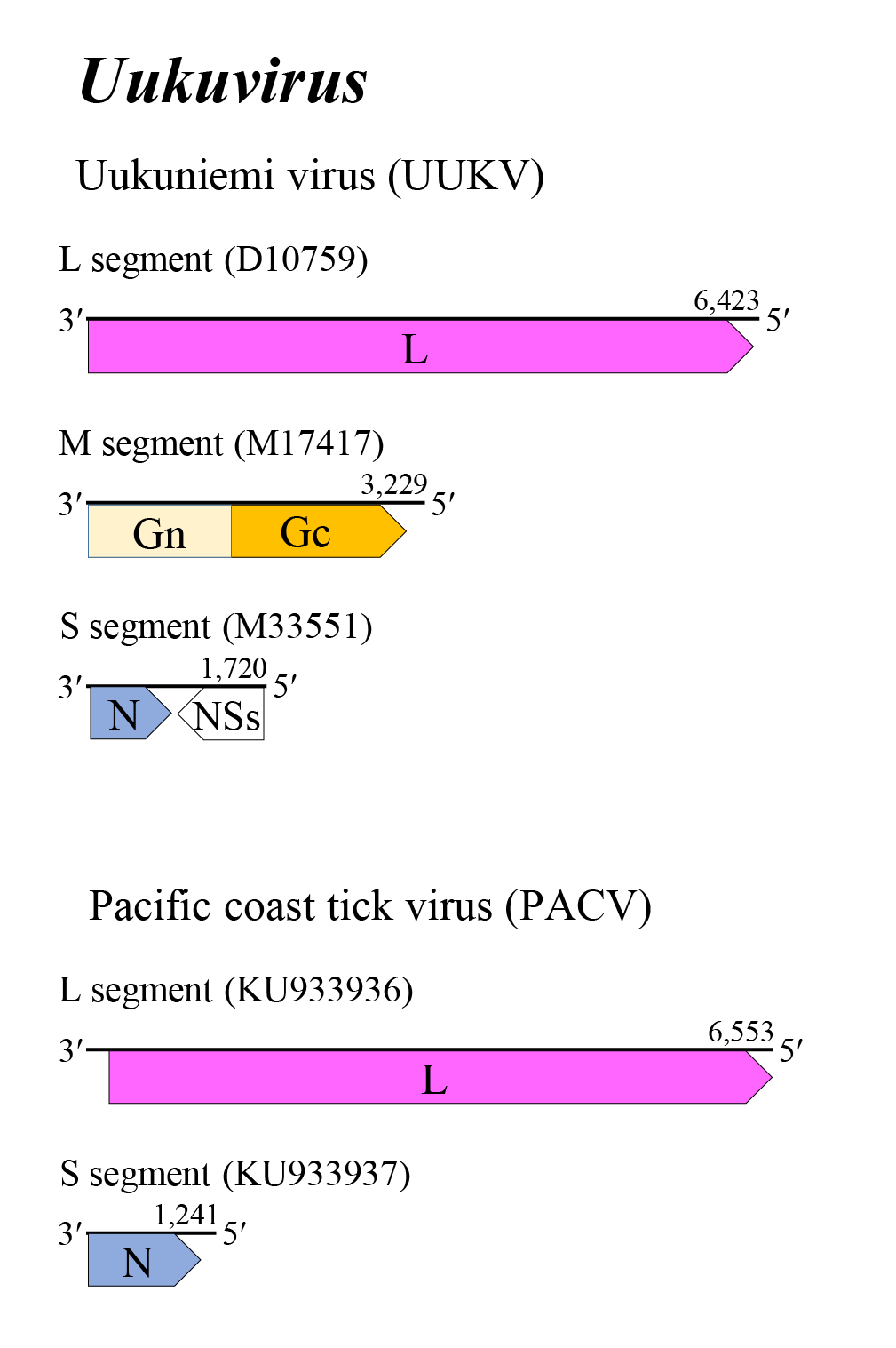 |
| Figure 1 Uukuvirus. Genome organization of some uukuviruses. Coloured boxes depict ORFs that encode N, nucleocapsid protein; Gn and Gc, external glycoproteins; and L, large protein. A white box depicts an ORF that encodes NSs, non-structural protein. |
Biology
Host range
Most uukuviruses infect mammals and birds and are transmitted by ticks. However, some uukuviruses replicate only in ticks. Hosts of uukuviruses include cattles [Bos taurus (Linnaeus, 1758)], common murres [Uria aalge (Pontoppidan, 1763)], reindeer (Rangifer tarandus Linnaeus), rodents [Acomys cahirinus (E. Geoffrey, 1803), A. cahirinus (E. Geoffrey, 1803), Aethomys hindei (Thomas, 1902), Lemniscomys striatus (Linnaeus, 1758), Mus striatus (Linnaeus, 1758), Myoprocta acouchy (Erxleben 1777), Neotoma micropus (Baird, 1885), and Proechimys guyannensis (Geoffroy, 1803)], and humans. In general, uukuviruses have not been considered to be of public health and agricultural significance, although antibodies to some uukuviruses have been detected in sera from humans, cattle, wild animals, reptiles and birds (Hubálek and Rudolf 2012, Palacios et al., 2013).
Transmission
Uukuviruses are transmitted by particular species of ticks in a circulative, propagative manner. The major vectors are Argas robertsi (Hoogstraal, Kaiser & Kohls, 1968), A. reflexus (Fabricius, 1794), Dermacentor marginatus (Sulzer, 1776), D. nuttalli (Olenev, 1928), D. occidentalis (Marx, 1892), D. variabilis (Say, 1821), Haemaphysalis flava (Neumann, 1897), H. hystricis (Supino, 1897), H. leporispalustris (Packard, 1869), H. longicornis (Neumann, 1901), Ixodes ricinus (Linnaeus, 1758), I. uriae (White, 1852), Rhipicephalus microplus (Canestrini, 1888), and R. pulchellus (Gerstacker, 1873). Transovarial and venereal transmission have been demonstrated for some uukuviruses in their arthropod vectors. In addition to transmission by arthropod vectors, mammals can become infected through contact with the blood or body fluids of infected animals. Several uukuviruses have been isolated from the blood of common murres [Uria aalge (Pontoppidan, 1763)] and from ticks collected from nests of cliff swallows (Petrochelidon pyrrhonota Vieillot); avian host and/or vector movements may result in virus dissemination (Palacios et al., 2013, Li et al., 2015, Matsuno et al., 2015, Ejiri et al., 2018).
Species demarcation criteria
The criteria demarcating species in the genus are:
• Less than 95% identity in the amino acid sequence of the L protein.
Relationships within the genus
Phylogenetic relationships across the genus have been established from maximum likelihood trees generated from L protein amino acid sequences (Figure 2 Uukuvirus).
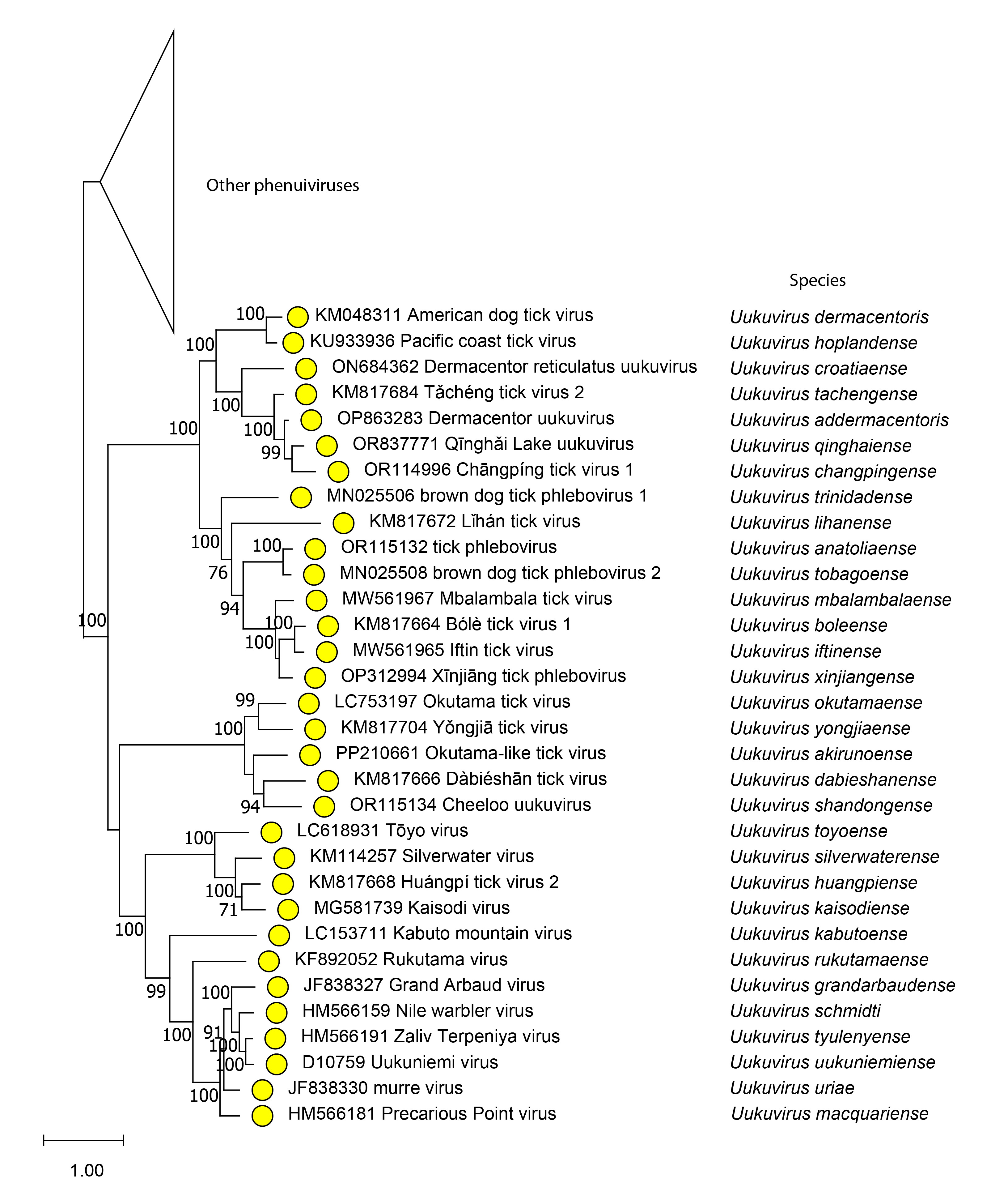 |
| Figure 2 Uukuvirus. Phylogenetic analysis of uukuvirus species. Phylogenetic reconstruction is based on a MAFFT-alignment of the RdRP amino acid sequences of phenuivirids using E-INS algorithm. The ML phylogenetic tree was inferred using RaxML-NG performing 1,000 bootstrap replicates. Trees were inferred under the WAG substitution model. Tree branches are proportional to genetic distances between sequences and the scale bars at the bottom indicate substitutions per amino acid. Full tree shown in Figure 3 Phenuiviridae. For alignment and treefile see Resources section. |
Related, unclassified viruses
| Virus name | Accession number | Virus abbreviation |
| Bangxi phenu tick virus 1 | L: ON746499 | BAPTV1 |
| brown dog tick phlebovirus 2_Tulcea47 | L: MW561138; S: MW561139* | BDTPV2_Tulcea47 |
| brown dog tick phlebovirus 2_Tulcea1 | L: MW561136; S: MW561137* | BDTPV2_Tulcea1 |
| Gissar virus | L: KJ425423*; M: KJ425424*; S: KJ425425 | GISV |
| Lanjan virus | L: PV804588; M: PV804589; S: PV804590 | LanV |
| Limansky tick phlebovirus | L: ON812423 | LTPV |
| Manawa virus | L: JQ924565*; M: JQ924566*; S: JQ924567* | MWAV |
| Qingdao tick uukuvirus | L: OQ513654; M: OQ513655; S: OQ513656 | QDTUV |
| soybean cyst nematode associated rice stripe virus | L: HM849041 | SCNaRSV |
| tick phlebovirus_TIGMIC-2 | L: ON812321 | TIPV_ |
| Tongren perib tick virus 2 | L: ON746493 | TPTV2 |
| Tongren Phenu tick virus 1 | L: ON746497 | TPTV1 |
| Toyo virus-TIGMIC_1 | L: ON812171 | TOYOV1 |
| Zhangjiakou phenu tick virus 1 | L: ON746500 | ZHPTV1 |
* Sequences do not comprise the complete genome segment.
Virus names and virus abbreviations are not official ICTV designations.