

Family: Phenuiviridae
Genus: Phasivirus
Distinguishing features
Phasiviruses were originally isolated from Culex mosquitoes in Australia. Seven viruses are assigned to the genus Phasivirus. The phasivirus genome has four genes, encoding a large protein (L), two external glycoproteins (Gn and Gc) and a nucleocapsid protein (N). Based on well-supported Maximum Likelihood or Maximum Clade Credibility trees inferred from complete L protein sequences, viruses classified in the genus Phasivirus form a monophyletic cluster clearly distinguished from other phenuivirids (Aguiar et al., 2015, Hobson-Peters et al., 2016, Shi et al., 2016, Shi et al., 2019, Williams et al., 2020).
Virion
Morphology
Virions are spherical or pleomorphic, 131.9 ±4.9 nm in diameter, and have surface glycoprotein projections which are embedded in a lipid bilayer envelope. Mature virions contain four major structural proteins of approximately 256, 56, 53 and 30 kDa that correspond to the RNA-directed RNA polymerase (RdRP), Gc, Gn, and N proteins of phenuivirids (Hobson-Peters et al., 2016).
Nucleic acid and Protein
The phasivirus genome encompasses three segments of negative-sense RNA. The terminal nucleotides of each segment occur in a canonical, conserved sequence (in coding sense) 5′-ACACAGAGAC…GUCUUUGUGU-3′ and may form the panhandle structures typical of other members of the class Bunyaviricetes (Table 2 Phenuiviridae). All three genomic RNAs contain untranslated regions flanking a single ORF which, based on comparisons with other negative-sense RNA viruses, is predicted to be contained in the virus-complementary strand. The L segment (6.7–7.0 kb) encodes a protein with a predicted molecular mass of 251–262 kDa that is homologous with the bunyaviral RdRP domain. The M segment (3.7–4.5 kb) encodes a protein of 128–155 kDa that is homologous with phlebovirus glycoprotein G1 and G2 sequences, as well as with the phlebovirus glycoprotein C-terminal Ig-like domain. The S segment (0.8–1.5 kb) encodes a protein of 29–50 kDa that is homologous with the tenuivirus/phlebovirus N domain (Table 3 Phenuiviridae) (Aguiar et al., 2015, Hobson-Peters et al., 2016, Shi et al., 2016, Shi et al., 2019, Williams et al., 2020).
Genome organization and replication
Phasivirus genome arrangement is similar to that of beidiviruses, citriciviruses, goukoviruses, hudiviruses, hudoviruses, pidchoviruses and tanzaviruses (Figure 1 Phasivirus). Three segments putatively encode L, the glycoprotein precursor (comprising Gn and Gc) and N, respectively. The Gn and Gc glycoproteins of 72–90 kDa and 53–56 kDa were earlier referred to as G1 and G2 based on apparent size following gel electrophoresis. The phasivirus genome does not encode either of the non-structural proteins NSs or NSm. Details of virus replication are unknown (Aguiar et al., 2015, Hobson-Peters et al., 2016, Shi et al., 2016, Shi et al., 2019, Williams et al., 2020).
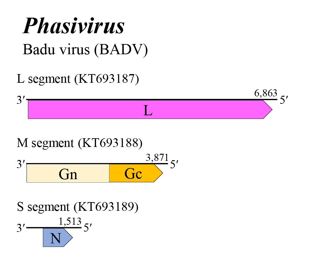 |
| Figure 1 Phasivirus. Genome organization of a phasivirus. Coloured boxes depict ORFs that encode N, nucleocapsid protein; Gn and Gc, external glycoproteins; and L, large protein. |
Biology
Phasivirus RNA has been found in the muscid fly Atherigona orientalis (Schiner, 1868) and in the mosquitoes Aedes aegypti (Linnaeus, 1762) and Culex quinquefasciatus (Say, 1823). Phasiviruses replicate in these insects but not in vertebrate cells (Aguiar et al., 2015, Hobson-Peters et al., 2016, Shi et al., 2016, Shi et al., 2019, Williams et al., 2020).
Species demarcation criteria
The criteria demarcating species in the genus are:
• Less than 95% identity in the amino acid sequence of the RdRP
Related, unclassified viruses
| Virus name | Accession number | Virus abbreviation |
| Hämeenlinna phasivirus | L: ON955138*; M: ON955139; S: ON955140 | HaPhasV |
| Broome phasivirus 1 | L: MT498819*; M: MT498821*; S: MT498820 | BrPhasV1 |
* Sequences do not comprise the complete genome segment.
Virus names and virus abbreviations are not official ICTV designations.