

Family: Peribunyaviridae
Genus: Khurdivirus
Distinguishing features
The first khurdivirus was isolated from coots in Astrakhan Oblast, Russia in 2001 (Galkina et al., 2005). Although the khurdivirus genome has complementary terminal ends identical to those found in orthobunyavirus genomes, khurdiviruses do not encode the non-structural M (NSm) protein that is encoded by all known orthobunyaviruses (Dimitry Konstantinovich et al., 2015).
Virion
Morphology
Mature virions are enveloped, spherical particles less than 220 nm in diameter (Galkina et al., 2005).
Nucleic acid
The khurdivirus genome consists of three segments of negative-sense RNA including the L, M, and S segments. The terminal nucleotides of each segment (S, M, and L) occur in a canonical, conserved sequence (in coding sense) 5′-AGTAGTGT…ACACTACT-3′, which is similar in sequence to those of members of the genera Orthobunyavirus and Lakivirus (Dimitry Konstantinovich et al., 2015).
Proteins
The proteins expressed form the khurdivirus genome include the large (L) protein with its RNA-directed RNA polymerase (RdRP), glycoprotein N (Gn), glycoprotein C (Gc), and the nucleoprotein (N). (Table 2 Peribunyaviridae). The Gc of khurdivirus is significantly shorter than the Gc of orthobunyaviruses with which it shares approximately 30% amino acid identity. These viruses do not encode any non-structural proteins (Dimitry Konstantinovich et al., 2015) (Figure 1.Khurdivirus).
Genome organization and replication
The khurdivirus genome is similar to the genomes of other peribunyaviruses (Figure 1 Khurdivirus). The S, M, and L segments encode the N, Gn and Gc, and L proteins, respectively. The L protein includes the six conserved RNA-directed RNA polymerase (RdRP) motifs including pre-A, and motifs A–E in the central region which shares 62% amino acid identity with bunyamwera virus (Dimitry Konstantinovich et al., 2015). The khurdivirus genome does not encode either of the non-structural proteins NSs or NSm (Dimitry Konstantinovich et al., 2015).
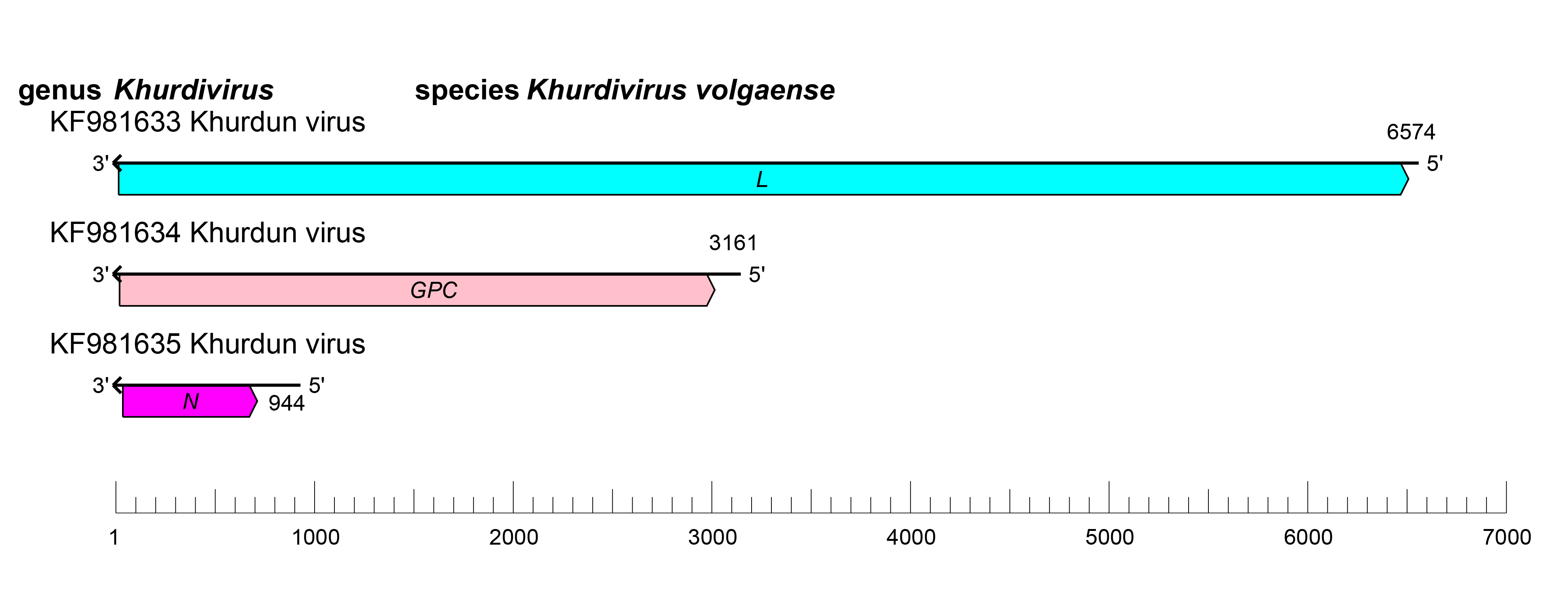 |
| Figure 1 Khurdivirus. Khurdivirus coding strategy. vcRNAs are depicted in 3'→5' direction and mRNAs are depicted in a 5′→3′ direction. Coloured boxes on the mRNAs depict ORFs that encode the N, nucleocapsid protein; Gn and Gc, external glycoproteins; L, large protein. |
Replication and morphogenesis in the Golgi are likely to be similar to that of other members of the family; however, this has not been thoroughly examined (see ‘Genome organization and replication’ on the family page).
Biology
Khurdun virus, the only member of the genus, has been isolated from coots (Fulica atra) in the Volga River delta (Al'khovskii et al., 2013). Currently, additional isolates of Khurdun virus from pygmy cormorants (Phalacrocorax pygmaues) have been described, but no other vertebrates or arthropod vectors have been associated with khurdiviruses (Dimitry Konstantinovich et al., 2015).
Antigenicity
Khurdiviruses are antigenically distinct from co-circulating orthobunyaviruses, but antigenic relationships within the genus are not described (Galkina et al., 2005).
Species demarcation criteria
Not defined as the genus currently includes only a single species.