

Family: Nudiviridae
Genus: Deltanudivirus
Distinguishing Features
The genus Deltanudivirus currently has only a single classified species, Deltanudivirus tipoleraceae. The virus (ToNV) was isolated in the 1950s from the cabbage crane fly Tipula oleracea [Diptera, Tipulidae] (Smith and Xeros 1954, Bézier et al., 2015). ToNV produces occlusion bodies with a size of 2−5 × 2 µm (Figure 1.Deltanudivirus), which are filled with enveloped, rod-shaped virions. Phylogenetic analysis using core genes (Figure 2.Nudiviridae) shows that ToNV is not closely related to any of the other nudiviruses, and therefore this species has been assigned to a separate genus. ToNV appears most closely related to the endogenous nudiviral gene clusters in Cotesia parasitic wasp genomes (Bézier et al., 2015), which are involved in bracovirus particle formation. ToNV, therefore, appears as a close relative of an ancient nudivirus that integrated into an ancestral parasitic wasp.
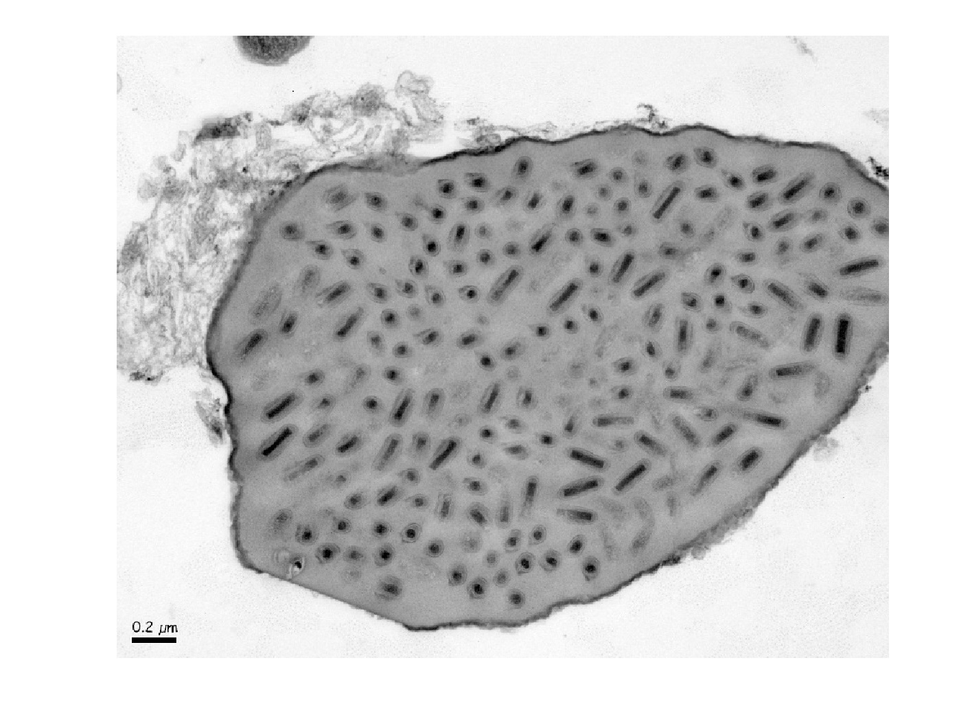 |
| Figure 1.Deltanudivirus. Transmission electron microscopy image of an ultrathin section of a Tipula oleracea nudivirus occlusion body (OB), in which the individual, enveloped virions are visible. The OBs were fixed in 4% paraformaldehyde and 1% glutaraldehyde in 0.1 M phosphate buffer (pH 7.2), and post-fixed in 2% osmium tetroxide, and embedded in Epon resin (Sigma). Ultrathin sections were stained with 5% uranyl acetate and 5% lead citrate then examined in a JEOL 1011 transmission electron microscope. Image: Annie Bézier and Julien Gaillard. |
Genome organisation and replication
The circular dsDNA genome sequence of ToNV comprises of 145 704 bp and contains five direct repeat regions and 131 predicted ORFs. Proteomic analysis clearly identified 48 different viral proteins in ToNV occlusion bodies, four others were detected based on a limited number of peptides (Bézier et al., 2017). It is not clear yet which gene encodes the occlusion body matrix protein.
Biology
ToNV was described as the causative agent of crane fly nucleopolyhedrosis. This disease is rarely observed which explains why there are relatively few data concerning its biology. The first report (Rennie 1923) described a viral disease characterized by the presence of moon-shaped occlusion bodies within the nuclei of fat tissue cells from Tipula paludosa (considered to be a baculovirus at that time). Later research showed that only hemocytes were infected (Smith and Xeros 1954). Subsequent work, described infected larvae from the Tipula genus with an increased volume of milky and slimy hemolymph containing irregular-shaped polyhedra; larvae showed a whitish coloration, decreased turgidity, reduced weight, diminished reactions to stimuli and an absence of pupation before dying (Meynardier et al., 1964). The occlusion bodies in T. paludosa were further analysed by Bergoin & Guelpa (Bergoin and Guelpa 1977) using EM studies. Bioassays conducted to test ToNV oral infectivity with an occlusion body solution (Meynardier et al., 1964, Bézier et al., 2015) confirmed that per os infection is a possible route for its transmission, as it is followed by the appearance of disease symptoms. It is not known whether the ToNV and the virus described in T. paludosa (earlier named as TpNPV based on (Rennie 1923)) belong to the same or different species.
Species demarcation criteria
Not defined as there is only one species in the genus.