

Family: Artoviridae
Ralf G. Dietzgen, Dàohóng Jiāng, Jens H. Kuhn, Arnfinn L. Økland, Nikos Vasilakis and and Gongyin Ye
The citation for this ICTV Report chapter is the summary published as Dietzgen et al., (2019):
ICTV Virus Taxonomy Profile: Artoviridae, Journal of General Virology, 100, 1202–1203.
Corresponding author: Ralf G. Dietzgen ([email protected])
Edited by: Jens H. Kuhn and Stuart G. Siddell
Posted: June 2019, updated June 2020, March 2022
PDF: ICTV_Artoviridae.pdf (2020 version)
Summary
The family Artoviridae was created in 2018 for the established monospecific genus Peropuvirus and six new species for invertebrate viruses all discovered by high throughput sequencing (Li et al., 2015, Shi et al., 2016). One new species for a plant virus (Yang et al., 2020) was added to this genus in 2022. A second genus, Hexartovirus, including two species for viruses of sea lice (Økland et al., 2019) and barnacles (Shi et al., 2016) respectively, was added in 2020. Artovirids form a family in the haploviricotine order Mononegavirales (Kuhn et al., 2021)
Table 1.Artoviridae. Characteristics of members of the family Artoviridae.
| Characteristic | Description |
| Example | Pteromalus puparum negative-strand RNA virus 1 (KX431032), species Peropuvirus pteromali |
| Virion | Enveloped, spherical particles, 100–130 nm in diameter |
| Genome | Negative-sense, unsegmented RNA of approximately 12 kb |
| Replication | Nuclear: the RNA-directed RNA polymerase engages with the ribonucleoprotein complex at the genome’s 3′-end |
| Translation | Individual, putatively polyadenylated, mRNAs are translated in the cytoplasm |
| Host Range | Barnacles, copepods, odonates, parasitoid wasps, pillworms, woodlice, plants (eggplant) |
| Taxonomy | Realm Riboviria, kingdom Orthornavirnae, phylum Negarnaviricota, subphylum Haploviricotina, class Monjiviricetes, order Mononegavirales. Genus Peropuvirus includes 9 species and genus Hexartovirus includes 4 species. |
Virion
Morphology
Virions of Pteromalus puparum negative-strand RNA virus 1 (PpNSRV-1) are enveloped and spherical with a diameter of 100–130 nm (Figure 1.Artoviridae). Virion morphology and structure is not available for other artovirids, which are only known from genomic sequence data.
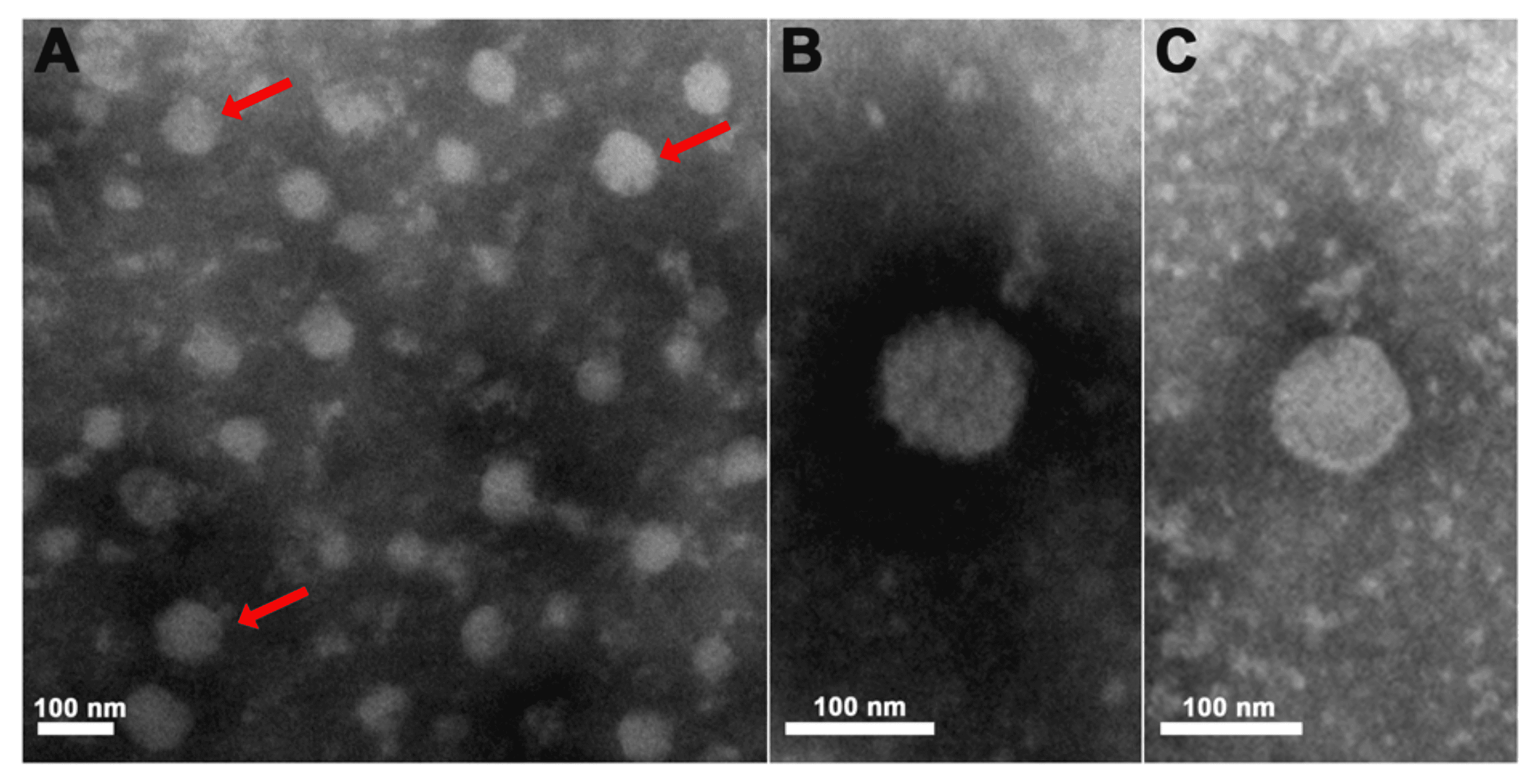 |
| Figure 1.Artoviridae. Electron micrographs of purified PpNSRV-1 particles from follicular cells. (A) Electron micrographs of purified PpNSRV-1 virions. (B) and (C) are the magnification pictures. Red arrows indicate viral particles. From (Wang et al., 2017), figure S3). |
Spherically shaped PpNSRV-1 particles are present in follicular cells of the ovaries of infected parasitoid wasps (Figure 1.Artoviridae). Similarly, virion-like particles stacked in intracellular vesicles have been observed in cells of the digestive tract. The spherical particles of PpNSRV-1 are similar to particles produced by nyamivirids (Wang et al., 2017, Dietzgen et al., 2017).
Physicochemical and physical properties
None reported.
Nucleic acid
Unsegmented, single-stranded RNA genome of 11–12.5 kb.
Proteins
Artovirids encode up to 5 structural proteins. Among them are the nucleocapsid protein (N), glycoprotein (G) and RNA-directed RNA polymerase (RdRP) domain-containing large protein (L) that are identified based on sequence similarity and structural properties shared with mononegaviral homologues. The functions of the other encoded proteins are largely unknown and likely are those of matrix and polymerase cofactor proteins.
The amino acid sequence of the PpNSRV-1 L protein is 26% identical to that of Midway virus (MIDWV, Nyamiviridae). The PpNSRV-1 ORF IV protein contains 7 potential O-linked and 4 potential N-linked glycosylation sites and 29 potential phosphorylation sites, suggesting that ORF IV may encode the viral glycoprotein G. The U1 protein is predicted to have 37 potential phosphorylation sites, whereas the U2 protein has 9 potential phosphorylation sites. The U3 protein contains 37 potential O-linked glycosylation sites. Thus, U3 may encode a glycosylated matrix protein M and U1 may encode the viral nucleoprotein N. Based on the conserved genomic organization of mononegaviruses, U2 is suggested to encode a phosphoprotein (P) (Wang et al., 2017).
Lipids
None reported.
Carbohydrates
None reported.
Genome organization and replication
Artovirid single-stranded RNA genomes are up to 12.5 kb (Figure 2.Artoviridae). All known artovirids have unsegmented genomes. The number of open reading frames (ORFs) varies. Genomes usually encode hypothetical proteins of unknown function (U1, U2 and U3), a putative glycoprotein (G) protein and a large protein (L) encoding an RNA-directed RNA polymerase domain: in the gene order 3'-U1-U2-U3-(G)-L-5'. The complete L gene sequences for all known members are known. Knowledge of artovirid replication is limited.
The PpNSRV-1 genome contains five large, independently-transcribed, non-overlapping ORFs flanked by 3′-leader and 5′-trailer regions whose terminal nucleotides do not exhibit obvious complementarity. Typical conserved transcription initiation and termination motifs are identified upstream and downstream, respectively, of each putative ORF.
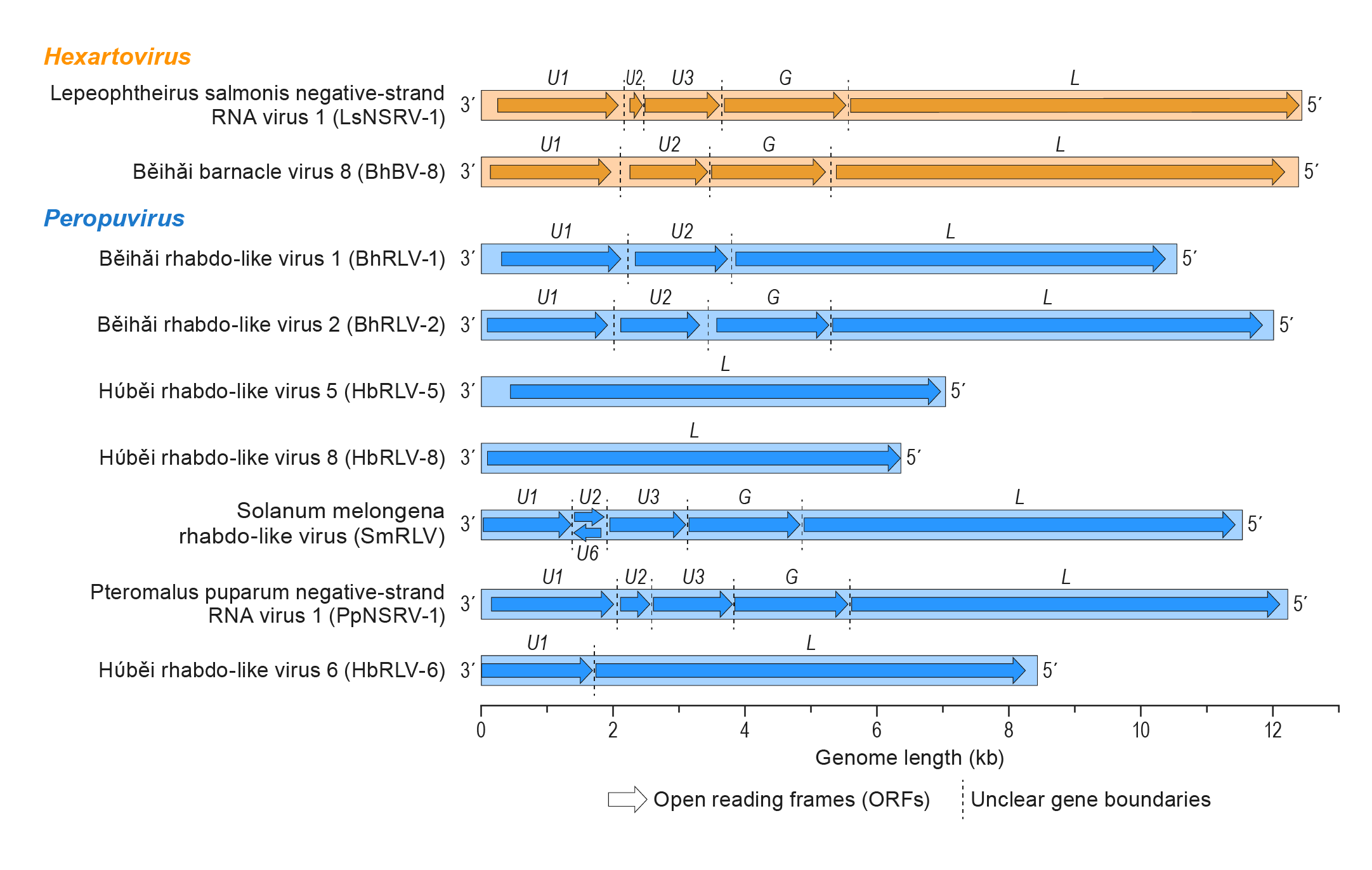 |
| Figure 2. Artoviridae. Artovirus genome organization. Some virus genome sequences may be incomplete. |
Biology
Members of the genus Peropuvirus infect parasitoid wasps, barnacles, pillworms, woodlice odonates, or copepods, and one has been identified in eggplant (Yang et al., 2020). Members of the genus Hexartovirus infect sea lice (copepods) or barnacles.
PpNSRV-1 was originally isolated from a laboratory parasitoid strain of a pteromalid wasp (Pteromalus puparum Linnaeus, 1758). PpNSRV-1 is present in various tissues and life stages of the parasitoid and is transmitted vertically through females and males. PpNSRV-1 increases adult longevity and impairs several fitness parameters of the wasp but has no influence on successful parasitism. PpNSRV-1 moderates the offspring sex ratio by decreasing female offspring numbers (Wang et al., 2017).
Lepeophtheirus salmonis negative-strand RNA virus 1 (LsNSRV-1) was detected in salmon lice (Lepeophtheirus salmonis Kroger, 1837) infecting farmed Atlantic salmon (Salmo salar Linnaeus, 1758) in Norway. LsNSRV-1 was present in various tissues of male and female salmon lice and was detected in all developmental stages. Viral RNA was detected in both the cytoplasm and nucleoli of infected cells (Økland et al., 2019).
Derivation of names
Artoviridae: from arthropod
Peropuvirus: from Pteromalus puparum, the species to which the pteromalid wasp belongs, from which the family’s typical member, PpNSRV-1, was first isolated.
Hexartovirus: from the crustacean class Hexanauplia and artovirus.
Relationships within the family
Phylogenetic relationships between the RdRP amino acid sequences of artovirids and members of related mononegavirus families are shown in Figure 3.Artoviridae.
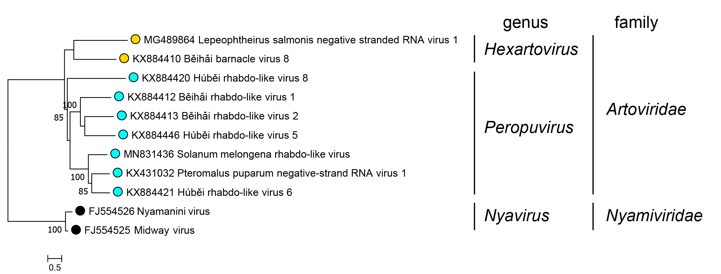 |
| Figure 3. Artoviridae. Phylogenetic maximum likelihood tree of L protein of members of the family Artoviridae. Artovirids are classified in the two genera Peropuvirus and Hexartovirus based on L protein amino acid sequences. Viruses of the two genera form monophyletic clades. Representative members of related mononegavirus families are included for comparison. The ML tree was produced using an LG + G + I model in MEGA10 (Kumar et al., 2018). This phylogenetic tree and corresponding sequence alignment are available to download from the Resources page. |
Relationships with other taxa
Members of the family Artoviridae are phylogenetically most closely related to members of the families Bornaviridae, Lispiviridae, Mymonaviridae, and Nyamiviridae (Figure 3.Artoviridae).