Genus: Rhizidiovirus
Chapter Version: ICTV Ninth Report; 2009 Taxonomy Release
Virion properties
Morphology
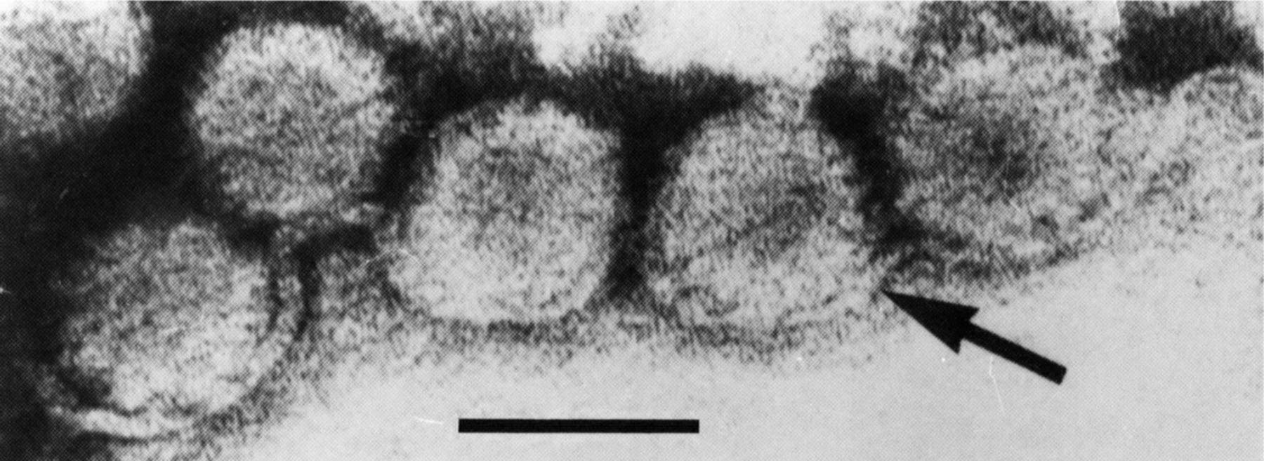
Virions are isometric, 60 nm in diameter (Figure 1).
Physicochemical and physical properties
The buoyant density of virions in CsCl is 1.31 g cm−3; S20,w is 625. Virions contain 10% nucleic acid.
Nucleic acid
Virions contain a single molecule of dsDNA with a Mr of 16.8 106 and a G+C ratio of 42%.
Proteins
Virions contain at least 14 polypeptides with sizes in the range of 26−84.5 kDa.
Lipids
None reported.
Carbohydrates
None reported.
Genome organization and replication
Particles appear first in the nucleus.
Biological properties
The virus appears to be transmitted in a latent form in the zoospores of its host Rhizidiomyces sp. Activation of the virus, which occurs under stress conditions such as heat, poor nutrition, or aging, results in cell lysis. Rhizidiomyces sp. is a water mold belonging to the kingdom Protista (Chromalveolata); it is no longer classified in the kingdom Fungi. Therefore, there are currently no recognized dsDNA viruses that naturally replicate in fungi.
No research has been done on this dsDNA virus system since 1983.
Species demarcation criteria in the genus
Not applicable.
List of species in the genus Rhizidiovirus
| Rhizidiomyces virus |
|
|
| Rhizidiomyces virus |
| (RZV) |
Species names are in italic script; names of isolates are in roman script. Sequence accession numbers [ ] and assigned abbreviations ( ) are also listed.
List of other related viruses which may be members of the genus Rhizidiovirus but have not been approved as species
None reported.
Similarity with other taxa
None reported.
Derivation of name
Rhizidio: from name of the host Rhizidiomyces sp.
Further reading
Dawe, V.H. and Kuhn, C.W. (1983). Virus-like particles in the aquatic fungus, Rhizidiomyces. Virology, 130, 10-20.
Dawe, V.H. and Kuhn, C.W. (1983). Isolation and characterization of a double-stranded DNA mycovirus infecting the aquatic fungus, Rhizidiomyces. Virology, 130, 21-28.
Contributed by
Ghabrial, S.A.
Figures
Figure 1 Negative contrast electron micrograph of particles of an isolate of Rhizidiomyces virus, which have been isolated from mechanically broken sporangia, are observed attached on a membrane-like structure (arrow). The bar represents 50 nm.
(From Dawe and Kuhn (1983). Virology, 130, 1020; with permission.)