Family: Corticoviridae
Chapter Version: ICTV Ninth Report; 2009 Taxonomy Release
Since only one genus is currently recognized, the family description corresponds to the genus description.
Genus Corticovirus
Type species Pseudoalteromonas phage PM2
Distinguishing features
PM2 is a virulent virus infecting gram-negative Pseudoalteromonas species. The characteristic feature of the corticovirus is the highly supercoiled circular double stranded DNA genome. The virions are composed of an icosahedral protein capsid and an inner protein-rich membrane enclosing the genome.
Virion properties
Morphology
Icosahedral virions consist of an internal membrane and an outer protein capsid that has a diameter of 57 nm between facets (Figure 1). The capsid consists of 200 major capsid protein P2 trimers that are organized on a pseudo T=21 lattice. Protein P2 is composed of two beta-barrels disposed normal to the capsid surface. The P2 trimers have pseudo-six-fold symmetry and the structure is stabilized by calcium ions. Spikes protrude about 8 nm from the capsid surface at the five-fold vertices. The spikes are homopentamers and formed of protein P1. P1 is composed of three beta-barrel domains arranged end to end. The distal C-terminal domains of P1 are used for receptor recognition. The N-termini of P1 form pentagonal assemblies at the vertices. The inner membrane (47 nm in diameter) contains host plasma membrane-derived phospholipids and virus-encoded proteins P3 to P10. Transmembrane proteins P3 and P6 are organized into a lattice on the membrane vesicle surface, on which the outer protein capsid is assembled.
Physicochemical and physical properties
The mass of the virion is about 45×106. The buoyant density in CsCl is 1.28 g cm−3 and in sucrose 1.26 g cm−3, and the S20,w is 293S. Virions are stable at pH 6–8, and are very sensitive to ether, chloroform and detergents. The virion stability is strongly dependent on sodium and calcium ions. Virions are unstable when frozen.
Nucleic acid
The genome is a highly supercoiled circular dsDNA of 10,079 bp (Mr of 6.6×10⁶). DNA comprises about 14% of the virion weight and the G+C content is 42.2%. The phage PM2 genome has been sequenced (AF155037).
Proteins
The genome has 21 putative genes, 17 of which have been shown to code for structural proteins (P1–P10) and nonstructural proteins (P12–P18; Table 1).
Table 1 Pseudoalteromonas phage PM2 proteins
| Proteina | Mass (kDa) | Location/functionb |
| P1 | 37.5 | Spike protein, receptor binding |
| P2 | 30.2 | Major capsid protein |
| P3 | 10.8 | Major membrane protein |
| P4 | 4.4 | Membrane |
| P5 | 17.9 | Membrane |
| P6 | 14.3 | Major membrane protein |
| P7 | 3.6 | Membrane |
| P8 | 7.3 | Membrane |
| P9 | 24.7 | Putative packaging ATPase |
| P10 | 29.0 | Membrane |
| P12 | 73.4 | Replication initiation protein (N) |
| P13 | 7.2 | Transcription factor (N) |
| P14 | 11.0 | Transcription factor (N) |
| P15 | 18.1 | Regulative function (N) |
| P16 | 10.3 | Regulative function (N) |
| P17 | 6.0 | Lysis factor (N) |
| P18 | 5.7 | Lysis factor (N) |
a P is for protein; Arabic numeral corresponds to the Roman numeral of the gene.
b N is for non-structural protein.
Lipids
Particles are about 14% lipid by weight. The membrane contains about 34% phosphatidyl ethanolamine and about 66% phosphatidyl glycerol and trace amounts of phosphatidic acid and acyl-phosphatidyl glycerol. The lipids are derived from the host plasma membrane, but their composition deviates from that of the host bacterium. Lipids form an internal membrane with virus-specific integral membrane proteins.
Carbohydrates
None reported.
Genome organization and replication
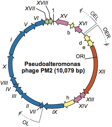
To infect and replicate, PM2 delivers its genome across the cell envelope of two known marine host strains: gram-negative Pseudoalteromonas species ER72M2 and BAL-31. Virions adsorb via the distal tips of the spike proteins to uncharacterized receptors. The internal membrane mediates the translocation of the supercoiled genome across the host cell envelopes via fusion. Replication of the PM2 genome, most probably by a rolling circle mechanism, takes place in proximity to the host cytoplasmic membrane. The genome is organized in three operons (Figure 2). Operons OEL and OER encode early function gene products: the replication initiation protein P12 and transcription regulatory proteins P13, P14, P15 and P16. Expression of the genes for structural proteins is under the control of the late promoter (OL), which is activated by the phage-encoded transcription factors P13 and P14. The release of mature virions from the cell occurs by a novel lysis mechanism.
Antigenic properties
Not known.
Biological properties
Phages are virulent and replicate in two known strains of marine host bacteria of the genus Pseudoalteromonas. Although PM2 is virulent and the sole member of the family Corticoviridae, using a comparative genomic approach, integrated corticoviral genetic elements have been identified to commonly reside within aquatic bacterial chromosomes.
Species demarcation criteria in the genus
Not applicable.
List of species in the genus Corticovirus
| Pseudoalteromonas phage PM2 |
|
|
| Pseudoalteromonas phage PM2 | [AF155037] | (PM2) |
Species names are in italic script; names of strains are in roman script. Sequence accession numbers [ ] and assigned abbreviations ( ) are also listed.
List of other related viruses which may be members of the genus Corticovirus but have not been approved as species
None reported.
List of unassigned species in the family Corticoviridae
None reported.
Phylogenetic relationships within the family
Not applicable.
Similarity with other taxa
Corticoviruses resemble tectiviruses in having a lipid bilayer underneath the isometric protein capsid. These viruses appear to differ by the genome organization and the infection mechanism, since no tectivirus-specific tail-like membrane tube is seen upon corticoviral infection. PM2 capsid architecture and capsid protein fold (trimeric protein with two beta-barrels forming hexagonal capsomers) have also been described in bacteriophages PRD1 and Bam35 (family Tectiviridae), the archaeal Sulfolobus turreted icosahedral virus (STIV; family not assigned), human adenovirus (family Adenoviridae), and in large eukaryotic viruses Paramecium bursaria Chlorella virus 1 (family Phycodnaviridae) and Chilo iridescent virus (family Iridoviridae).
Derivation of name
Cortico: from Latin cortex, “crust”, “bark”.
Further reading
Abrescia, N.G., Grimes, J.M., Kivelä, H.M, Assenberg, R., Sutton, G.C., Butcher, S.J., Bamford, J.K.H., Bamford, D.H. and Stuart, D.I. (2008). Insights into virus evolution and membrane biogenesis from the structure of the marine lipid-containing bacteriophage PM2. Mol. Cell, 31, 749-761.
Bamford, J.K.H. and Bamford, D.H. (2006). Lipid-containing bacteriophage PM2, the type organism of Corticoviridae. In: R. Calendar (Ed.), The Bacteriophages. Oxford University Press, New York, pp. 171-174.
Krupovi, M. and Bamford, D.H. (2007). Putative prophages related to lytic tailless marine dsDNA phage PM2 are widespread in the genomes of aquatic bacteria. BMC Genomics, 8, 236.
Männistö, R.H., Kivelä, H.M., Paulin, L., Bamford, D.H. and Bamford, J.K.H. (1999). The complete genome sequence of PM2, the first lipid-containing bacterial virus to be isolated. Virology, 262, 355-363.
Contributed by
Oksanen, H.M and Bamford, J.K.H.
Figures
Figure 1 (Left) X-ray crystallographic structure of a virion of Pseudoalteromonas phage PM2 at 7 resolution, viewed along two-fold axis of symmetry (courtesy of N.G.A. Abrescia). (Middle) A schematic presentation and (right) negative stain electron micrograph of Pseudoalteromonas phage PM2 particles. The bar represents 50 nm.
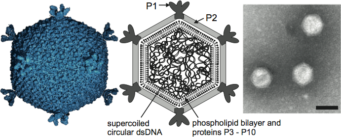
Figure 2 Genome organization of Pseudoalteromonas phage PM2 (PM2). The genome is a 10,079 bp highly supercoiled circular dsDNA molecule containing 21 ORFs (as arrows showing their orientations) and three operons (OER, OEL and OL). ORFs shown to code for functional proteins are classified as genes and given a Roman numeral. The different colors indicate the ORFs (yellow), a gene for replication initiation protein (orange) and the following groups of genes: transcriptional regulation (magenta), structural proteins (blue) and lysis (green).