
Family: Tospoviridae

Family: Tospoviridae
Paolo Margaria, Scott Adkins, Ozgur Batuman, Amalendu Ghosh, Holly R. Hughes, Rayapati A. Naidu, Massimo Turina and Anna E. Whitfield
The citation for this ICTV Report chapter is the summary to be published as Margaria et al., (2026): ICTV Virus Taxonomy Profile: Tospoviridae 2026, Journal of General Virology, (in press)
Corresponding author: Paolo Margaria ([email protected])
Edited by: Luisa Rubino and Evelien Adriaenssens
Posted: September 2026
Summary
Tospoviridae is a family of plant viruses with a genome consisting of three molecules of negative-sense or ambisense single-stranded (ss)RNA, designated S (about 2.9 kb), M (about 4.8 kb), and L (about 8.9 kb) (Table 1 Tospoviridae). Virions are enveloped, spherical or pleomorphic in morphology, 80–120 nm in size, with surface projections composed of two viral glycoproteins. The core of the virus particle contains ribonucleoproteins composed of the ssRNA components encapsidated by the nucleocapsid protein and a few copies of the viral RNA-directed RNA polymerase. The genome encodes two non-structural proteins. The family includes a single genus, Orthotospovirus. Members of the family are transmitted in nature by thrips (Thysanoptera: Thripidae) in a persistent, propagative manner. A broad host range and ability to infect major food and ornamental crops make these viruses threats to agricultural production wherever the virus and thrips vector(s) occur.
Table 1 Tospoviridae. Characteristics of members of the family Tospoviridae
| Characteristic | Description |
| Example | tomato spotted wilt virus (L: D10066; M: S48091; S: D00645), species Orthotospovirus tomatomaculae |
| Virion | Enveloped, round to pleomorphic virions with a diameter of 80–120 nm |
| Genome | Three linear, negative-sense or ambisense ssRNA molecules ranging from about 2.9 to about 8.9 kb in length, comprising about 17 kb in total |
| Replication | Cytoplasmic |
| Translation | From capped mRNAs that lack a poly(A) tail |
| Host range | Plants and thrips (Thysanoptera: Thripidae) |
| Taxonomy | Realm Riboviria, kingdom Orthornavirae, phylum Negarnaviricota, subphylum Polyploviricotina, class Bunyaviricetes, order Elliovirales: one genus including 36 species |
Virion
Morphology
Mature virions are spherical or pleomorphic, 80–120 nm in diameter, and display surface glycoprotein projections of 5–10 nm, which are embedded in a host-derived lipid bilayer envelope (Figure 1 Tospoviridae). The core of the virion contains ribonucleoprotein (RNP) complexes that consist of the viral genomic RNAs bound to the nucleocapsid protein (N) and a few molecules of the viral RNA-directed RNA polymerase (RdRP, also referred to as L protein). In the canonical view, each virion packages one copy of each genomic segment; however, de facto, virions differ in genomic content and only reach a reproducible genetic composition at the viral population scale, resembling the genome formula of multipartite viruses (Yvon et al., 2023). Dense masses of nucleocapsid material may accumulate in the cytoplasm; these masses may be composed of defective particles (Resende et al., 1991, Resende et al., 1992).
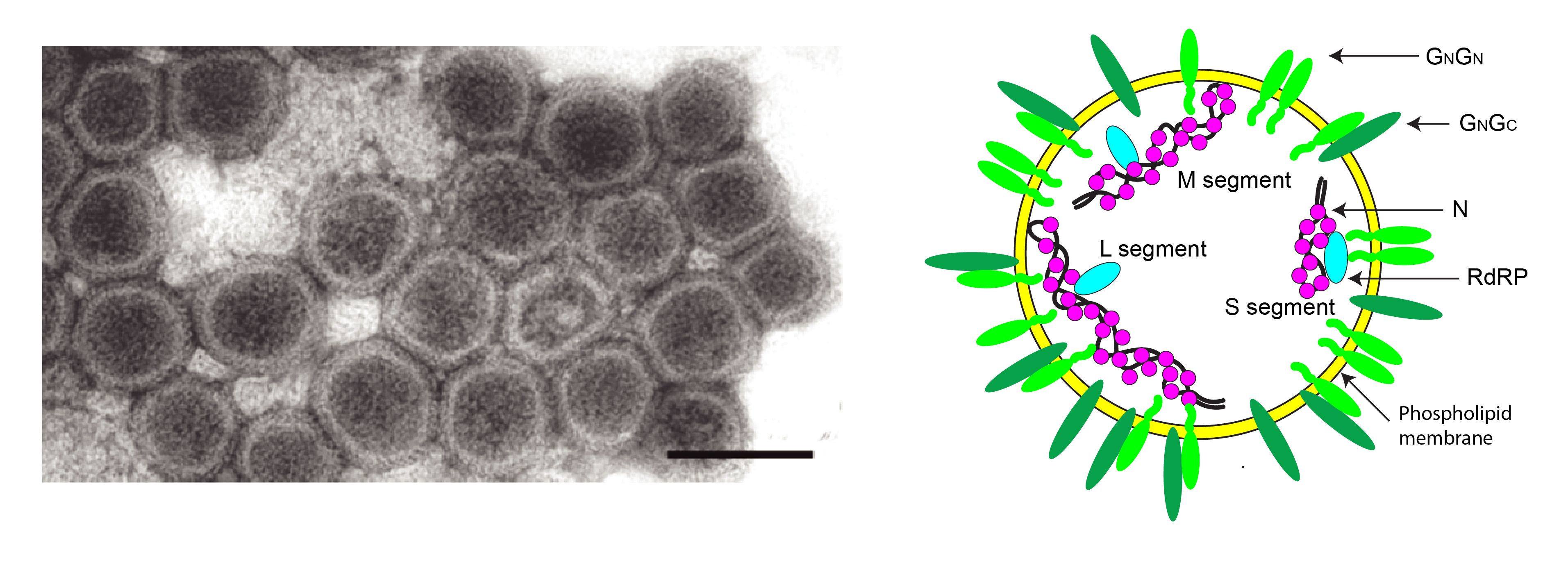 |
| Figure 1 Tospoviridae. Virion structure of tospovirids. (Left) Electron micrograph of negatively-stained particles of tomato spotted wilt virus. The bar represents 100 nm. (Courtesy of Dr Jan van Lent.). (Right) Virion graphical representation (N - nucleoprotein, RdRP – RNA-directed RNA polymerase, GN, GC – N-terminal and C-terminal glycoproteins, shown as monomers, heterodimers or homodimers). |
Physicochemical and physical properties
Virion composition is 5% nucleic acid, 70% protein, 5% carbohydrate and 20% lipid (Adkins 2000). Virion buoyant density in CsCl is 1.20 g/cm3 (Dijkstra and de Jager 1998).
Nucleic acid
The genome consists of three linear, single-stranded (ss)RNA molecules, that according to size are denoted as large (L, about 8.9 kb), medium (M, about 4.8 kb), and small (S, about 2.9 kb) RNA (de Haan et al., 1990, de Haan et al., 1991, Kormelink et al., 1992a). The L RNA is of negative polarity, while M and S RNA contain an ambisense gene arrangement, with two non-overlapping reading frames (ORFs) on opposite strands, separated by a non-coding intergenic region (IR). The termini of each of the RNA segments consist of an eight-nucleotide sequence (5′-AGAGCAAU-3′) that tends to be strictly conserved among members of different species in the family. Non-canonical octanucleotide sequences have been reported (Maneechoat et al., 2024). The remaining untranslated regions at the termini also exhibit a high degree of complementarity, facilitating base pairing of the RNA termini and the formation of panhandle structures, making genomic RNAs appear pseudocircular. Viral mRNAs lack a 3′-poly(A) tail and have heterogenous non-viral nucleotides at the 5′-terminus derived from host-cell mRNAs via a cap-snatching mechanism, resulting in a 5′-methylated cap leader fragment (Kormelink et al., 1992b, van Poelwijk et al., 1996).
Proteins
Virions of members of the family contain four structural proteins: a large protein (L; about 330 kDa), encoded by the L segment; the GN and GC glycoproteins, embedded in the phospholipid envelope and derived by proteolytic processing of a glycoprotein precursor (GPC; about 128 kDa), encoded by the M segment; and the nucleocapsid protein (N; about 29 kDa), encoded by the S segment.
Lipids
Virions are composed of a phospholipid membrane that is derived from host cell membranes where virions mature (Kikkert et al., 1999). This envelope is essential for virion integrity, glycoprotein anchoring, and transmission by insect vectors.
Carbohydrates
Virions contain 5% carbohydrate by weight (Adkins 2000).
Genome organization and replication
The genome organization and expression strategy for members of the family is depicted in Figure 2 Tospoviridae. The L segment is a negative-sense RNA and encodes on the viral-complementary (vc) strand the L protein, which functions as the viral RNA-directed RNA polymerase (RdRP) (de Haan et al., 1991, Adkins et al., 1995). Both the M and S RNA have an ambisense gene arrangement (de Haan et al., 1990, Kormelink et al., 1992a). The M segment encodes the glycoprotein precursor in negative orientation, which is thereafter processed into two glycoproteins (GN and GC, so designated based on the position relative to the amino and carboxy termini of the polyprotein) by proteolytic cleavage, most likely by ER-resident proteases, and a non-structural protein (NSm, about 33 kDa) on the viral strand. The S segment encodes the structural N protein on the vc-strand, along with a non-structural protein (NSs, about 52 kDa) on the viral strand. The GN and GC proteins are embedded in the virion lipid bilayer and are instrumental in the acquisition and transmission by the thrips vector (Whitfield et al., 2004, Whitfield et al., 2008). The NSm protein represents the viral movement protein, enabling the intercellular movement of infectious RNPs via a tubule-guided manner through plasmodesmata (Kormelink et al., 1994, Storms et al., 1995, Lewandowski and Adkins 2005). The N protein is the nucleocapsid protein; it assembles into stable trimers (Guo et al., 2017, Komoda et al., 2017) and encapsidates the genomic RNAs to form the RNP complexes. NSs acts as suppressor of the antiviral RNA-interference (RNAi) defense response in plants, and can bind both long and short dsRNA, with diverse affinities across Orthotospovirus species (Schnettler et al., 2010). In virulent isolates, NSs is highly expressed and may form paracrystalline or filamentous inclusions in infected plant cells (Kormelink et al., 1991, Kitajima et al., 1992). A functional NSs protein also appears to be needed for persistent infection and transmission by the vector, likely by suppressing antiviral RNAi within thrips (Margaria et al., 2014).
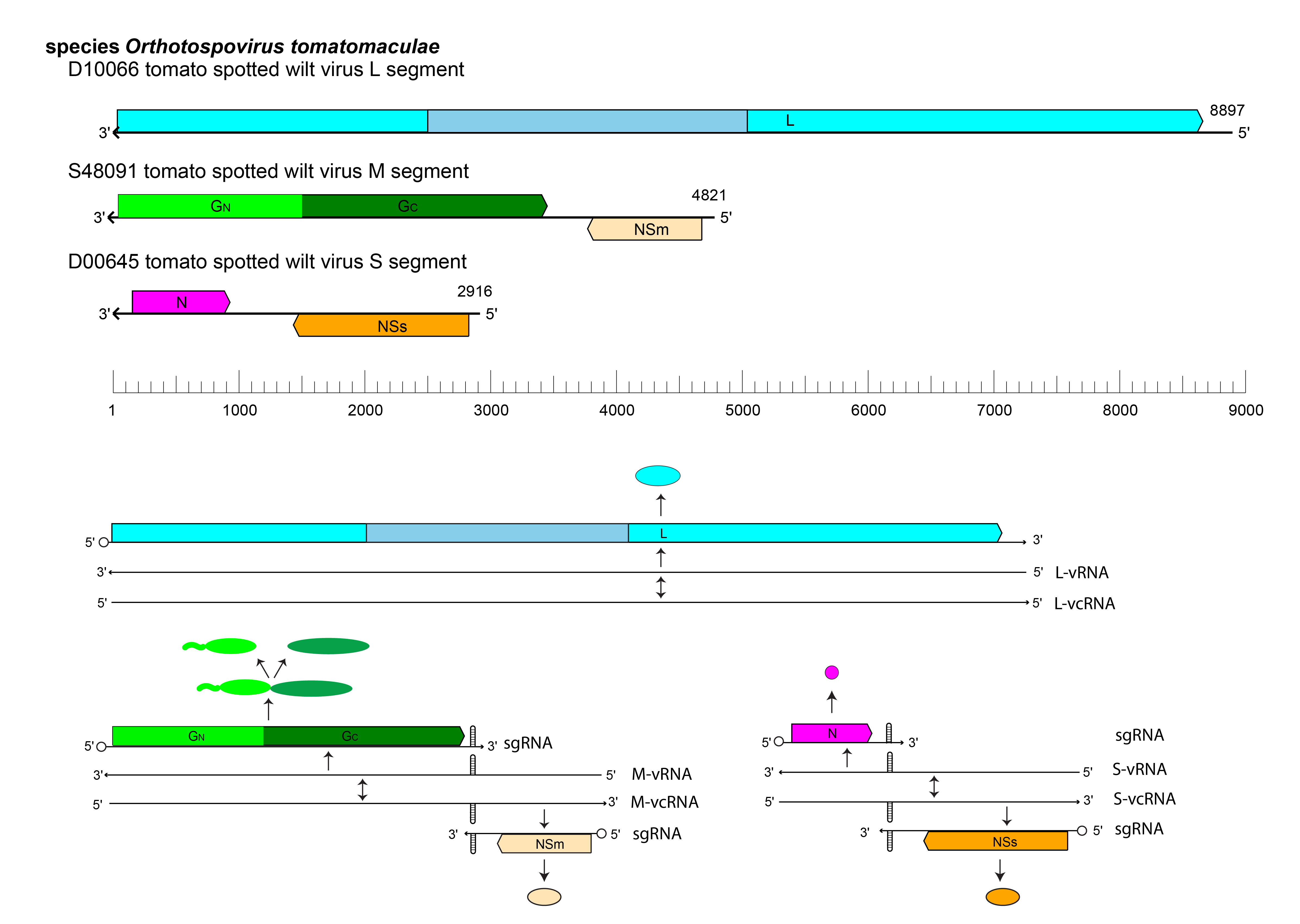 |
| Figure 2 Tospoviridae. Tospovirid genome organization and replication strategy. (Top) Genome organisation of tomato spotted wilt virus (TSWV), a representative member of the family Tospoviridae. Coloured boxes depict ORFs that encode L, large protein, with the central RNA-directed RNA polymerase motif indicated by darker shading; GN, envelope glycoprotein from the N-terminus of the glycoprotein precursor; GC, envelope glycoprotein from the C-terminus of the glycoprotein precursor; NSm: non-structural protein on the M segment; N, nucleocapsid protein; NSs: non-structural protein on the S segment. (Bottom) Schematic of orthotospovirus replication and expression. An open circle indicates a 5′-terminal cap. Hairpin structures are present in the intergenic regions of the M and S RNAs. vRNA: viral RNA; vcRNA: viral-complementary RNA; sgRNA: subgenomic RNA. |
All proteins encoded by the RNA genome are expressed from near-genome length (L protein) or subgenomic length mRNAs (GPC, NSm, N, NSs) that are transcribed from the genomic RNAs. Viral mRNAs are generated through a cap-snatching mechanism whereby the viral transcriptase complex cleaves mature host cellular mRNAs approximately 12–18 nucleotides downstream of the 5′ cap and utilizes the resulting capped oligonucleotides as primers for transcription on the viral RNA template (Kormelink et al., 1992b). Transcription termination of subgenomic mRNAs derived from the M and S RNA segments occurs within the intergenic region (IR), an A- and U-rich sequence predicted to form a stable hairpin structure that likely functions as a transcriptional terminator (van Knippenberg et al., 2005). The very short IR of the S segment of Polygonum ringspot virus (species Orthotospovirus polygonianuli) lacks the potential for formation of the predicted hairpin structure, supporting the existence of other means for the achievement of transcription termination of subgenomic RNAs (Ciuffo et al., 2008).
Replication of the viral RNA genome takes place in electron-dense cytoplasmic inclusions (viroplasms). Newly synthesized RNP complexes either traffic intra- and intercellularly, or mature into virus particles by acquisition of a lipid envelope at the Golgi complex through a process involving coordinated multimeric interactions among the three major structural proteins (N, GN, and GC) (Ribeiro et al., 2008, Ribeiro et al., 2009). During this process, RNP complexes are enwrapped by entire Golgi stacks, resulting in the formation of doubly enveloped virus particles that subsequently undergo membrane fusion events to generate mature, singly enveloped virions, which accumulate within large intracellular vesicles (Kikkert et al., 1999). From these reservoirs, virions are acquired by thrips during feeding on infected plants, thereby enabling virus transmission and dissemination.
Orthotospoviruses also replicate in specific tissues of infected thrips vectors (Ullman et al., 1992, Ullman et al., 1993, Wijkamp et al., 1993, Montero-Astúa et al., 2016). The rate of viral replication is key in influencing transmission efficiency and determining vector competence (Nagata et al., 2002, Rotenberg et al., 2009, Margaria et al., 2014).
Biology
At least 19 species of thrips (order Thysanoptera) from eight genera viz. Frankliniella, Thrips, Scirtothrips, Taeniothrips, Neohydatothrips, Dycothrips, Microcephalothrips and Ceratothripoides transmit orthotospoviruses (Table 2 Tospoviridae). Virus acquisition by thrips solely occurs during the larval stages after which the virus is passed transstadially to the adult. Acquisition rates decrease as larval thrips develop (Van De Wetering et al., 1996). Once acquired from infected plant cells, the virus needs to reach the midgut epithelial cells, and travel across several host membranes (Ullman et al., 1992, Nagata et al., 1999, de Assis Filho et al., 2002, Nagata et al., 2002, Whitfield et al., 2005, Montero-Astúa et al., 2016), until reaching the salivary glands. Adults, and partly second-instar larvae, can transmit the virus by the injection of viruliferous saliva into plant tissues.
Mechanical transmission via homogenized leaf tissue from infected plants is possible in experimental conditions.
Members of the family can infect a large number of plant species, including economically important vegetable crops such as tomato, bean, cucurbit, lettuce, pepper, potato, and soybean, various ornamental species, as well as numerous weed species. Tomato spotted wilt virus (TSWV, species Orthotospovirus tomatomaculae) has a very broad host range and a worldwide distribution, whereas other orthotospoviruses are much more limited in host range and geographic occurrence.
The best way to limit/prevent orthotospovirus-induced diseases involves management strategies that include virus resistance. Against TSWV, dominant resistance genes (Sw‑5b gene in tomato, the Tsw gene in pepper) are widely employed (Turina et al., 2016). Interaction between the TSWV NSm movement protein with SW-5 in tomato, and the NSs protein with Tsw in pepper, lead to induction of the resistance response (Jahn et al., 2000, Hoffmann et al., 2001, Margaria et al., 2007, de Ronde et al., 2014, Zhu et al., 2017). The continuous use of resistant cultivars has resulted in the onset of TSWV resistance-breaking (RB) isolates worldwide (Black et al., 1995, Roggero et al., 2002, Aramburu and Marti 2003, Margaria et al., 2004, Sharman and Persley 2006, Margaria et al., 2007, Batuman et al., 2017, Fidan and Sarı 2019, Macedo et al., 2019, Yoon et al., 2021, Almási et al., 2023, Chinnaiah et al., 2023, Gautam et al., 2023, Lahre et al., 2023, Macedo et al., 2024), including ‘double RB’ isolates (Chinnaiah et al., 2026, Forgia et al., 2026). Although the Sw-5b gene confers resistance to a few other American clade orthotospoviruses, RB emergence has thus far occurred only with TSWV.
Reassortment among isolates within a species appears to be a common evolutionary strategy (Qiu et al., 1998, Qiu and Moyer 1999, Tentchev et al., 2011, Margaria et al., 2015, Adegbola et al., 2019, Peng et al., 2025); interspecific reassortment can occur (Webster et al., 2015, Silva et al., 2019). Reassortment, within-segment recombination and selection have all contributed to the origin and diversification of orthotospoviruses (Tentchev et al., 2011, Butković et al., 2021).
Table 2 Tospoviridae. Vectors associated with the transmission of members of the family Tospoviridae
Species | Virus name | Vector |
Orthotospovirus arachinecrosis | groundnut bud necrosis virus | Frankliniella schultzei, Scirtothrips dorsalis, Thrips palmi, T. parvispinus |
Orthotospovirus arachianuli | groundnut ringspot virus | Frankliniella gemina, F. occidentalis, F. schultzei, F. intonsa |
Orthotospovirus arachiflavamaculae | groundnut yellow spot virus | Scirtothrips dorsalis |
Orthotospovirus impatiensnecromaculae | Impatiens necrotic spot virus | Frankliniella occidentalis. F. intosa, F. schultzei, F. fusca |
Orthotospovirus iridimaculaflavi | iris yellow spot virus | Thrips tabaci, Frankliniella fusca |
Orthotospovirus polygonianuli | Polygonum ringspot virus | Dictyothrips betae |
Orthotospovirus tomatoflavi | tomato chlorotic spot virus | Frankliniella occidentalis, F. schultzei, F. intonsa |
Orthotospovirus tomatomaculae | tomato spotted wilt virus | Frankliniella bispinosa, F. cephalica, F. gemina, F. fusca, F. intonsa, F. occidentalis, F. schultzei, Thrips setosus, T. tabaci |
Orthotospovirus citrullonecrosis | watermelon bud necrosis virus | Thrips palmi |
Orthotospovirus citrullomaculosi | watermelon silver mottle virus | Thrips palmi |
Orthotospovirus cucurbichlorosis | zucchini lethal chlorosis virus | Frankliniella zucchini |
Orthotospovirus alstroemeriflavi | Alstroemeria yellow spot virus | Thrips tabaci |
Orthotospovirus alstroemerinecrosis | Alstroemeria necrotic streak virus | Frankliniella occidentalis |
Orthotospovirus arachiflavi | groundnut chlorotic fan-spot virus | Scirtothrips dorsalis |
Orthotospovirus callaflavi | calla lily chlorotic spot virus | Thrips palmi |
Orthotospovirus capsiciflavi | Capsicum chlorosis virus | Ceratothripoides claratris, Thrips palmi, Frankliniella schultzei, Microcephalothrips abdominalis |
Orthotospovirus capsicimaculaflavi | pepper chlorotic spot virus | unknown |
Orthotospovirus chrysanthinecrocaulis | chrysanthemum stem necrosis virus | Frankliniella occidentalis, F. gemina, F. schultzei, F. intonsa |
Orthotospovirus eustomae | lisianthus necrotic ringspot virus | unknown |
Orthotospovirus fatsiae | Fatsia japonica ringspot-associated virus | unknown |
Orthotospovirus glycininecrovenae | soybean vein necrosis virus | Neohydatothrips variabilis, Frakliniella fusca, F. schultzei, F. tritici |
Orthotospovirus hippeflavi | Hippeastrum chlorotic ringspot virus | Taeniothrips eucharii |
Orthotospovirus meloflavi | melon yellow spot virus | Thrips palmi |
Orthotospovirus melotessellati | melon severe mosaic virus | unknown |
Orthotospovirus morivenae | mulberry vein banding-associated virus | unknown |
Orthotospovirus phaseolinecrotessellati | bean necrotic mosaic virus | unknown |
Orthotospovirus tomatanuli | tomato yellow ring virus | Thrips tabaci, Microcephalothrips abdominalis |
Orthotospovirus tomatozonae | tomato zonate spot virus | Frankliniella occidentalis |
Orthotospovirus tomato necroanuli | tomato necrotic ringspot virus | Thrips palmi, Ceratothripoides claratris |
Orthotospovirus limonii | Limonium orthotospovirus 1 | unknown |
Orthotospovirus barlerichlorosis | Barleria chlorosis-associated virus | unknown |
Orthotospovirus scadoxiflavianuli | Scadoxus chlorotic ringspot virus | unknown |
Orthotospovirus mercurialis | Mercurialis orthotospovirus 1 | unknown |
Orthotospovirus macadamianuli | Macadamia ringspot-associated virus | unknown |
Orthotospovirus capsiciflavianuli | chilli yellow ringspot virus | unknown |
Orthotospovirus tomatonecromaculae | tomato necrotic spot-associated virus | unknown |
Antigenicity
Antigenic variation among species is primarily driven by differences in the nucleocapsid and glycoprotein proteins, supporting serogroup classification (Adam et al., 1995).
Derivation of names
Tospoviridae: from tomato spotted wilt virus; the suffix -viridae for family taxa
Orthotospovirus: from the Ancient Greek ὀρθός (orthos), meaning “correct” or “straight” and tospovirus; the suffix -virus for genus taxa
Demarcation criteria for taxa within the family
Genus demarcation criteria are not currently defined as there is only one genus (Orthotospovirus) in the family. Based on aspects that consider phylogeny and biological properties, the possibility of creating multiple genera within the family Tospoviridae should be considered in a future taxonomic proposal (Peng et al., 2014, Oliver and Whitfield 2016, Butković et al., 2021).
Relationships within the family
Phylogenetic analyses using N protein sequences groups members of the family Tospoviridae into at least five distinct phylogenetic clades (Oliver and Whitfield 2016, Butković et al., 2021).
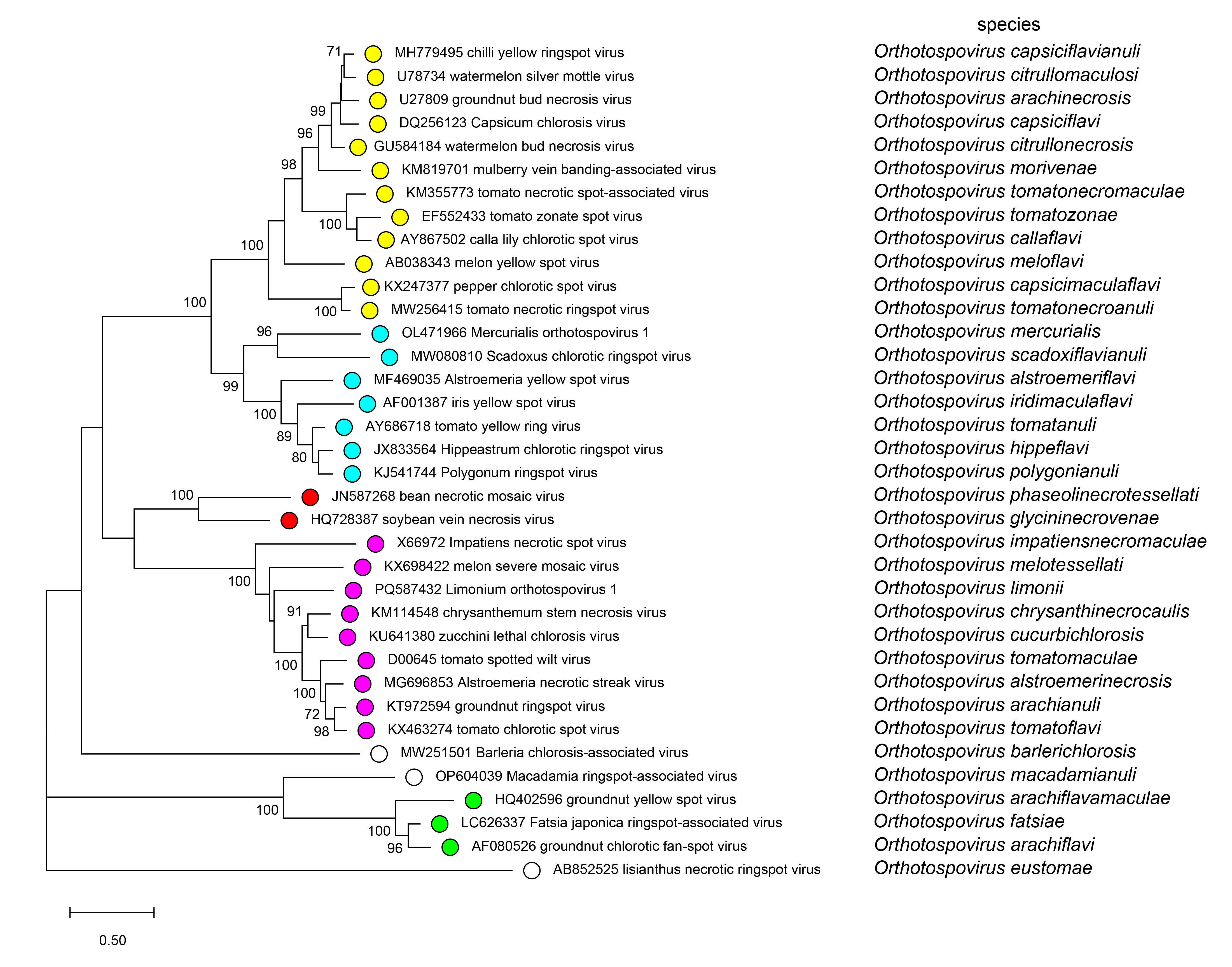 |
| Figure 3 Tospoviridae. Phylogenetic tree of orthotospovirus N protein sequences. The evolutionary history was inferred by using the Maximum Likelihood method and JTT matrix-based model (Jones et al., 1992) with 320 positions in the final dataset using MEGA11 (Tamura et al., 2021). The tree with the highest log likelihood (-11958.77) is shown. Bootstrap support is indicated where this was > 70%. A discrete Gamma distribution was used to model evolutionary rate differences among sites (4 categories (+G, parameter = 1.4970)) with 1.61% of sites invariable. The scale indicates number of substitutions per site. Tips are coloured according to clades as follows: yellow – watermelon silver mottle virus clade, cyan – iris yellow spot virus clade, red – bean necrotic mosaic virus clade, magenta – tomato spotted wilt virus clade, green – groundnut yellow spot virus clade, no colour – monotypic clades for Barleria chlorosis-associated virus, Macadamia ringspot-associated virus and lisianthus necrotic ringspot virus. |
Relationships with other taxa
The family Tospoviridae belongs to the class Bunyaviricetes, which currently includes 16 families of negative-sense segmented RNA viruses infecting arthropods, protozoans, plants, and animals (Kuhn et al., 2024). Three families within the class (Fimoviridae, Phenuiviridae, and Tospoviridae) include viruses that infect plants as primary host. Plant viruses in the families Tospoviridae and Fimoviridae share a closer relationship than with plant-infecting members of the family Phenuiviridae which are in a different order (Hareavirales) in the class Bunyaviricetes (Kormelink et al., 2021, Kuhn et al., 2024).