
Family: Tymoviridae (Interim Report)

Family: Tymoviridae (Interim Report)
This is a summary page created by the ICTV Report Editors using information from associated Taxonomic Proposals and the Master Species List.
Edited by: Luisa Rubino
Posted: November 2026
Summary
The family Tymoviridae includes ssRNA(+) viruses of plants (Table 1 Tymoviridae). The family Tymoviridae was established in 2002 (Master Species List #20).
Table 1 Tymoviridae. Characteristics of members of the family Tymoviridae.
Characteristic | Description |
Example | turnip yellow mosaic virus (X07441)), species Tymovirus brassicae |
Virion | Non-enveloped, icosahedral, 30 nm diameter |
Genome | ssRNA(+), 1 linear segment, 6.0–7.6 kb |
Replication | Membrane-associated replication complexes |
Translation | Cytoplasmic, translation from genomic RNA followed by polyprotein proteolytic processing and from subgenomic mRNAs |
Host range | Plants |
Taxonomy | Realm Riboviria, kingdom Orthornavirae, phylum Kitrinoviricota, class Alsuviricetes, order Tymovirales: 3 genera, 43 species |
Virion
Proteins
Tymovirid virions are isometric, ca. 30 nm in diameter, non-enveloped, with a prominent surface structure (Figure 1A Tymoviridae). The turnip yellow mosaic virus (TYMV) particle has 180 capsid protein subunits arranged in T=3 icosahedral symmetry (Figure 1B Tymoviridae). The capsid protein has a single jelly roll fold motif. The C-termini of all subunits are on the exterior, while the N-termini appear to be on the interior of the virus particle (Canady et al., 1996).
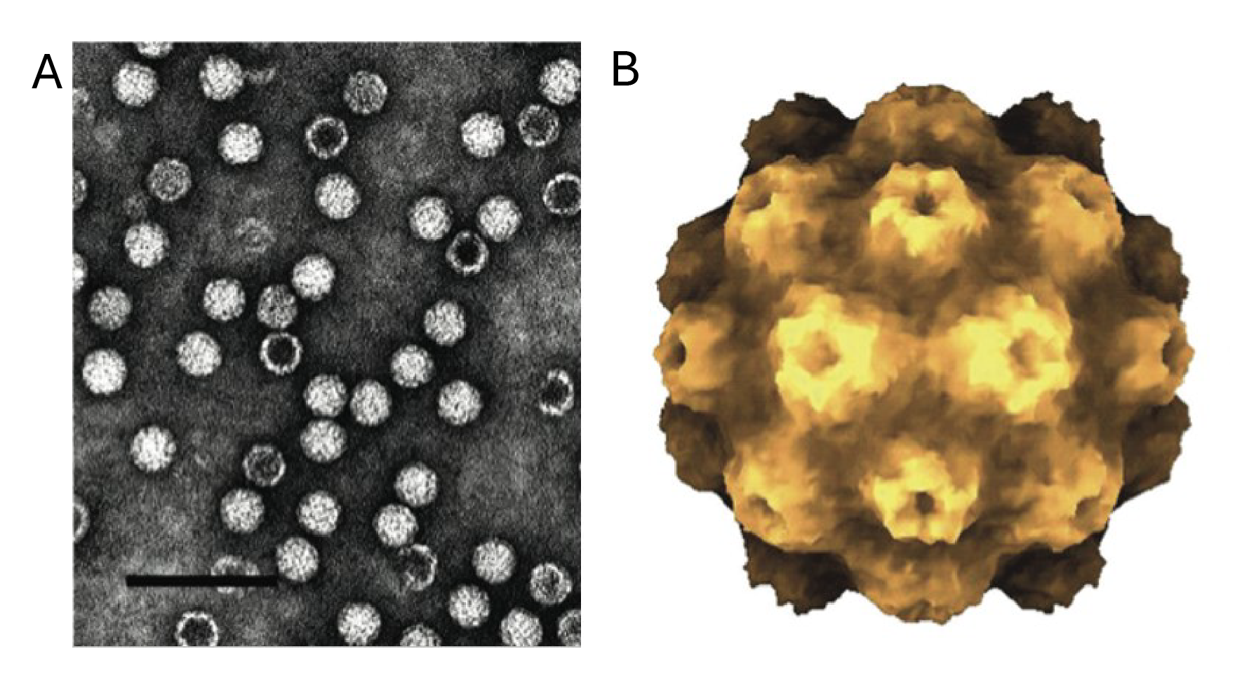 |
| Figure 1 Tymoviridae. A. Negative-contrast electron micrograph of virions and empty particles of belladonna mottle virus. The bar represents 100 nm. B. Atomic rendering of a virion of turnip yellow mosaic virus (Canady et al., 1996). |
Nucleic acid
The genome of tymovirids comprises a single, linear segment of positive-sense RNA of 6.0–7.6 kb. The genome of TYMV (and probably all tymovirids) has a 5′-m7GpppG cap structure. The 3′-terminus of tymovirid genomic RNA may be a tRNA-like structure, a poly(A) tract or atypical. The genome composition is C-rich and G-poor (Dreher 2004, Tzanetakis et al., 2009).
Genome organization and replication
Tymovirid genomes encode two proteins: the replicase protein (REP), with motifs indicative of methyltransferase, papain-like cysteine protease, helicase and RNA-directed RNA polymerase (RdRP) activities and a capsid protein (Figure 2 Tymoviridae). Tymovirus and some marafivirus genomes also encode an additional movement protein that may act as a weak suppressor of the plant RNA silencing response. The sequences encoding these proteins may be arranged as overlapping or non-overlapping ORFs and their expression may involve different combinations of proteolytic processing or translation from subgenomic mRNAs (Kadaré et al., 1992, Sabanadzovic et al., 2001, Edwards and Weiland 2014).
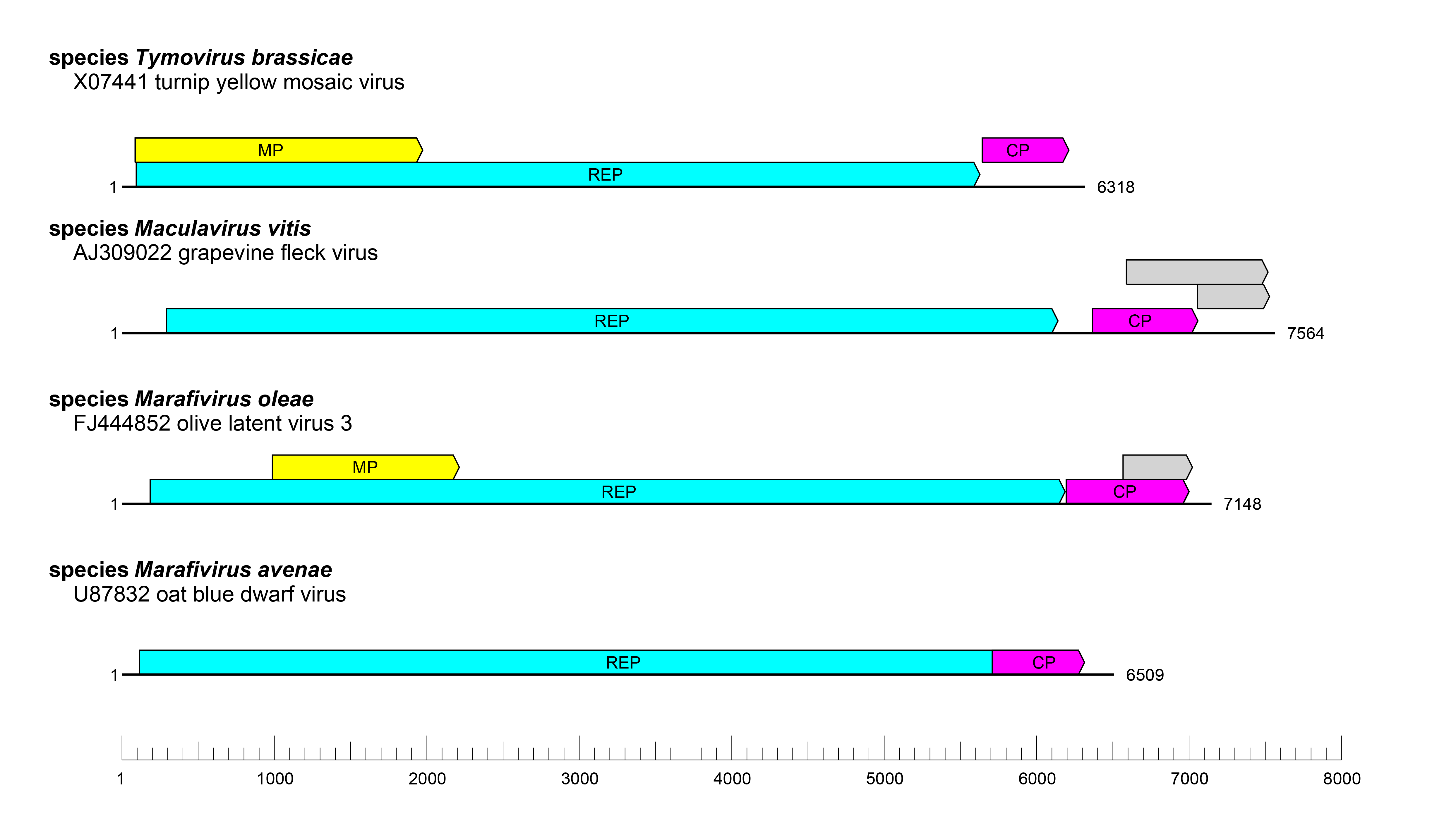 |
| Figure 2 Tymoviridae. Genome organisation of representative members of the family Tymoviridae. Boxes indicate open reading frames as annotated on the corresponding GenBank accession. Colours indicate coding regions for replicase (cyan), capsid (magenta), movement (yellow) and hypothetical (grey) proteins. |
Biology
Tymovirids are found world-wide and generally infect dicotyledonous plants, with the exception of some marafiviruses that primarily infect plants in the family Gramineae (Dreher 2012). Metagenomic analyses have detected unclassified tymovirid-like viruses in samples obtained from mosquitoes (Wang et al., 2012), bees and mites (de Miranda et al., 2015), and fungi (Li et al., 2016). Some members of the family are transmitted mechanically or by insect vectors, depending on the genus. In infected cells, tymovirid replication induces the derangement of chloroplasts (TYMV) or mitochondria (GFkV), undergoing peripheral vesiculation (Hatta et al., 1973, Martelli et al., 2002).
Demarcation criteria
The criteria demarcating genera are physicochemical and molecular characters combined with biological, epidemiological and cytopathological properties (Dreher 2012). The criteria demarcating species are genome sequence identity of less than 80%, capsid protein sequences less than 90% identical, differential host range, differences in the genome 3′-terminal structure, the number of ORFs, serological specificity and vector specificity, depending on the genus (Dreher 2012).
Phylogenetic analysis of the tymovirid replicase, RdRP core domain and CP proteins (for example, (Haenni 2008, Dreher 2012, Gibbs et al., 2026)) are generally consistent with the current classifications (Figure 3 Tymoviridae).
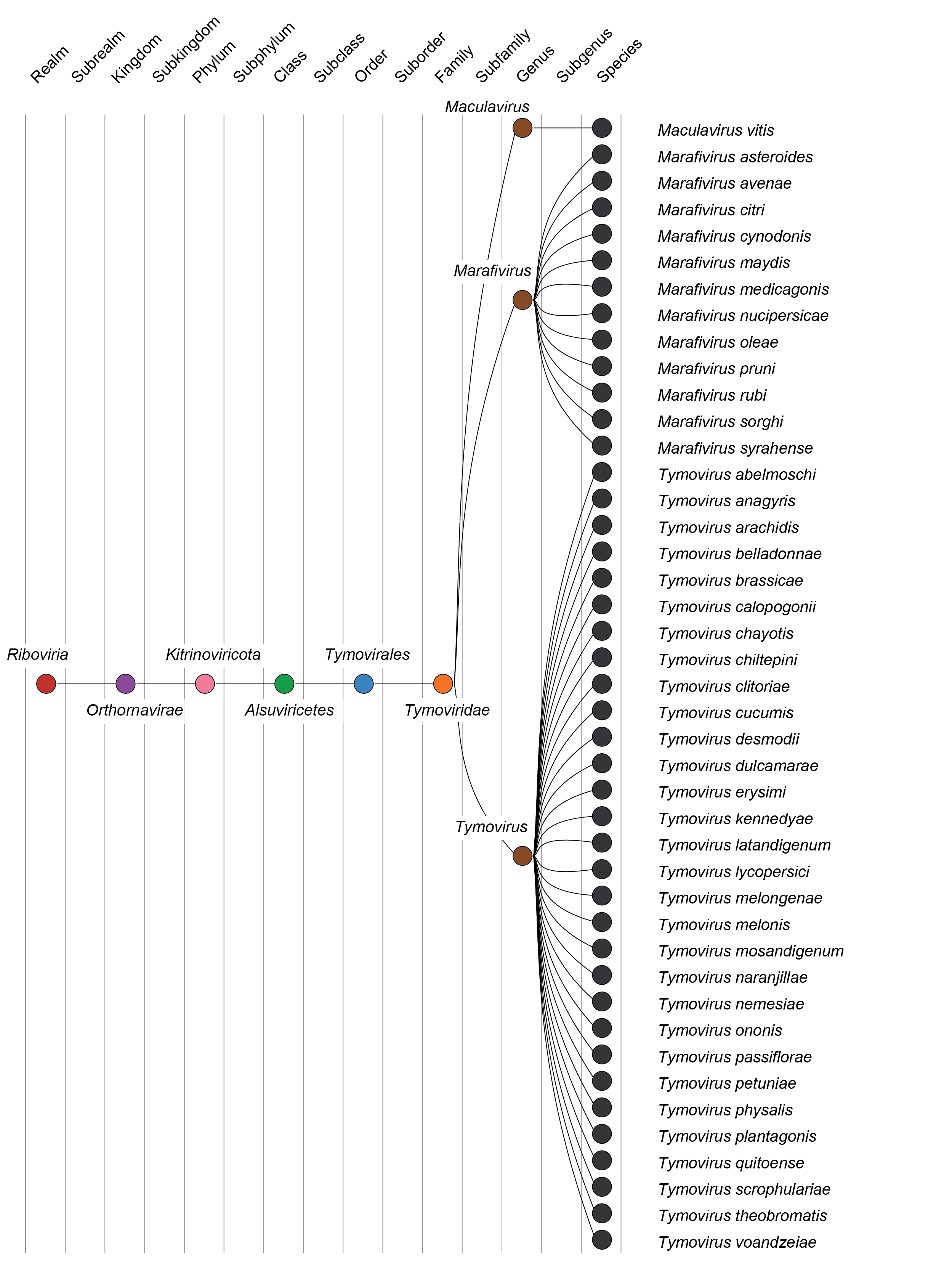 |
| Figure 3 Tymoviridae. Taxonomy of the family Tymoviridae. |
Derivation of names
Maculavirus: from the Latin macula, meaning fleck, referencing the characteristic "flecking" symptoms on the leaves of grapevines infected with grapevine fleck virus
Marafivirus: from the previous genus name Maize rayado fino virus group, itself derived from the virus name maize rayado fino virus
Tymoviridae, Tymovirus: from turnip yellow mosaic virus, a member of the family; the suffix -viridae for family taxa or -virus for genus taxa
References: Tymoviridae
Canady, M. A., S. B. Larson, J. Day and A. McPherson (1996). Crystal structure of turnip yellow mosaic virus. Nat Struct Biol 3: 771-81. [PubMed]
de Miranda, J. R., R. S. Cornman, J. D. Evans, E. Semberg, N. Haddad, P. Neumann and L. Gauthier (2015). Genome characterization, prevalence and distribution of a macula-like virus from Apis mellifera and Varroa destructor. Viruses 7: 3586-602. [PubMed]
Dreher, T. W. (2004). Turnip yellow mosaic virus: transfer RNA mimicry, chloroplasts and a C-rich genome. Mol Plant Pathol 5: 367-75. [PubMed]
Dreher, T. W., Edwards, M.C., Gibbs, A.J., Haenni, A.-L., Hammond, R.W., Jupin, I., Koenig, R., Sabanadzovic, S. and Martelli, G.P. (2012). Family Tymoviridae In Virus Taxonomy, the 9th Report of the International Committee on Taxonomy of Viruses, pp. 944-52. Edited by M. J. A. A. M. Q. King, E. B. Carstens & E. J. Lefkowitz. San Diego: Elsevier.
Edwards, M. C. and J. J. Weiland (2014). Coat protein expression strategy of oat blue dwarf virus. Virology 450-451: 290-6. [PubMed]
Gibbs, A. J., S. Fuentes, I. P. Adams, M. Hajizadeh, K. Ben Mansour, P. L. Guy, C. E. Fribourg, H. Ziebell, J. Kreuze, A. Fox and R. A. C. Jones (2026). A phylogeny of the tymoviruses, sensu stricto, and its global interpretation in space and time. Plant Dis 110: 300-13. [PubMed]
Haenni, A.-L., Dreher, T.W. (2008). Tymoviruses. In Encyclopedia of Virology (Third Edition), pp. 199–207. Edited by B. W. J. Mahy, Van Regenmortel, M.H.V. . Oxford: Academic Press.
Hatta, T., S. Bullivant and R. E. Matthews (1973). Fine structure of vesicles induced in chloroplasts of Chinese cabbage leaves by infection with turnip yellow mosaic virus. J Gen Virol 20: 37-50. [PubMed]
Kadaré, G., G. Drugeon, H. S. Savithri and A. L. Haenni (1992). Comparison of the strategies of expression of five tymovirus RNAs by in vitro translation studies. J Gen Virol 73: 493-8. [PubMed]
Li, P., Y. Lin, H. Zhang, S. Wang, D. Qiu and L. Guo (2016). Molecular characterization of a novel mycovirus of the family Tymoviridae isolated from the plant pathogenic fungus Fusarium graminearum. Virology 489: 86-94. [PubMed]
Martelli, G. P., S. Sabanadzovic, N. Abou Ghanem-Sabanadzovic and P. Saldarelli (2002). Maculavirus, a new genus of plant viruses. Arch Virol 147: 1847-53. [PubMed]
Sabanadzovic, S., N. A. Ghanem-Sabanadzovic, P. Saldarelli and G. P. Martelli (2001). Complete nucleotide sequence and genome organization of Grapevine fleck virus. J Gen Virol 82: 2009-15. [PubMed]
Tzanetakis, I. E., C. H. Tsai, R. R. Martin and T. W. Dreher (2009). A tymovirus with an atypical 3'-UTR illuminates the possibilities for 3'-UTR evolution. Virology 392: 238-45. [PubMed]
Wang, L., X. Lv, Y. Zhai, S. Fu, D. Wang, S. Rayner, Q. Tang and G. Liang (2012). Genomic characterization of a novel virus of the family Tymoviridae isolated from mosquitoes. PLoS One 7: e39845. [PubMed]