
Family: Polymycoviridae
Family: Polymycoviridae
Poliane Alfenas-Zerbini, Ioly Kotta-Loizou, Cauê Neves de Oliveira and Robert H. A. Coutts
The citation for this ICTV Report chapter is the summary to be published as Alfenas-Zerbini et al., (2026) Journal of General Virology ICTV Virus Taxonomy Profile: Polymycoviridae 2026 (in press)
Corresponding authors: Poliane Alfenas-Zerbini ([email protected]) and Ioly Kotta-Loizou ([email protected])
Edited by: Sead Sabanadzovic, Peter Simmonds and Evelien Adriaenssens
Posted: March 2022, updated May 2024, revised November 2026
Summary
Polymycoviridae is a family of viruses with multi-segmented non-conventionally encapsidated double-stranded (ds) RNA genomes (Table 1 Polymycoviridae). Typically, polymycovirids have four genomic segments, although some have up to eight. Each genomic segment is 0.7–2.5 kbp in length, infect fungi (ascomycetes and basidiomycetes) and oomycetes. The family currently includes three genera, Polymycovirus, Multimycovirus and Plurimycovirus, encompassing a total of 28 species.
Table 1 Polymycoviridae. Characteristics of members of the family Polymycoviridae.
| Characteristic | Description |
| Example | Aspergillus fumigatus tetramycovirus 1 (dsRNA1: HG975302; dsRNA2: HG975303; dsRNA3: HG975304; dsRNA4: HG975303), species Polymycovirus aspergilli |
| Virion | dsRNA associated with a viral protein, forming ribonucleoprotein complexes or filamentous particles |
| Genome | A total of 7.5–12.5 kbp of dsRNA in a multipartite genome (usually four segments, up to eight) |
| Replication | Both dsRNA and single-stranded (ss) RNA can be isolated from infected fungal hosts. Virions accumulate in the cytoplasm |
| Translation | From positive-sense (+) transcripts of genomic dsRNAs |
| Host range | Fungi (ascomycetes and basidiomycetes), oomycetes |
| Taxonomy | Realm Riboviria, kingdom, Orthornaviriae, phylum Pisuviricota; order Xenadelphovirales; three genera, Polymycovirus (9 species), Multimycovirus (17 species) and Plurimycovirus (2 species) |
Virion
Morphology
The majority of polymycoviruses do not form classical capsid-based virions. Instead, their genomic dsRNA is associated with a viral protein, forming ribonucleoprotein complexes rather than true capsids (Kanhayuwa et al., 2015, Zhai et al., 2016, Kotta-Loizou and Coutts 2017, Niu et al., 2018). Four members of the family have been reported to form filamentous particles, including Colletotrichum camelliae filamentous virus 1 (CcFV1), Pseudopestalotiopsis camelliae-sinensis polymycovirus 1, and Phyllosticta capitalensis polymycovirus 1 and 2. (Jia et al., 2017, Han et al., 2025), ranging from 10–34 nm in width and 950–1,500 nm in length (Figure 1 Polymycoviridae).
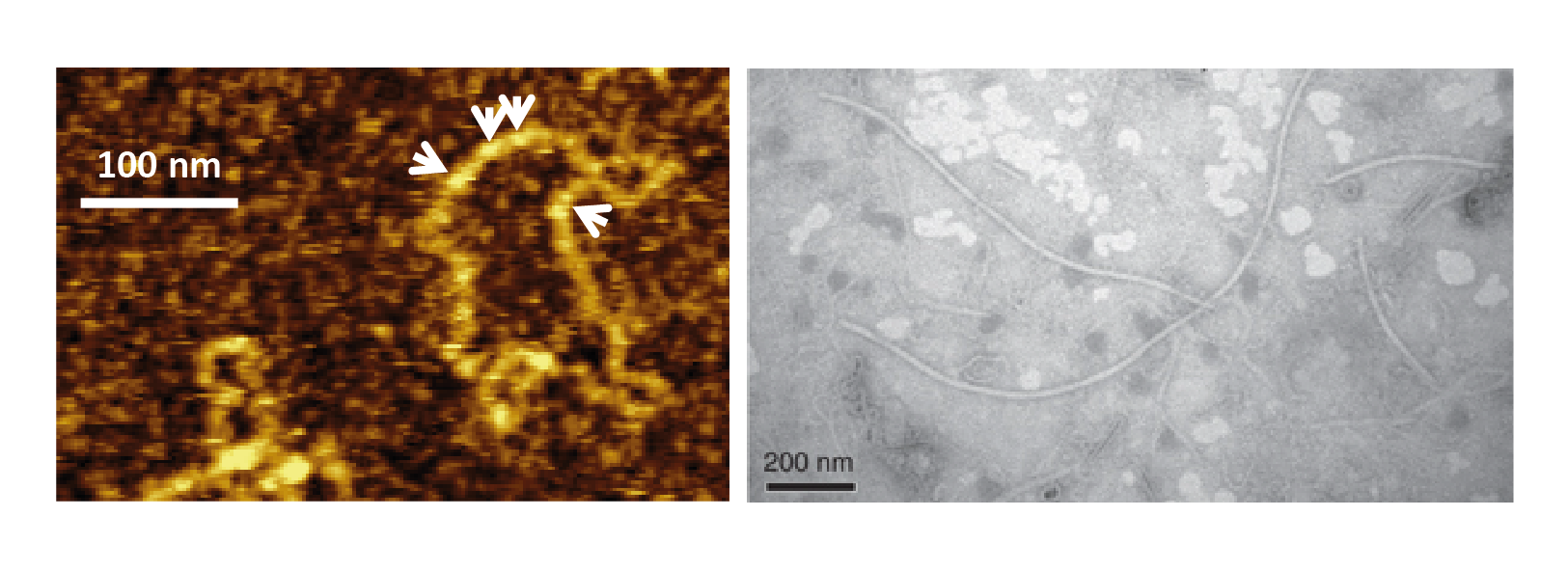 |
| Figure 1 Polymycoviridae. Polymycovirus virions. (Left) Visualisation using atomic force microscopy (AFM) of purified Aspergillus fumigatus tetramycovirus 1 ribonucleoprotein complexes, in which the dsRNA genome is coated by a viral protein rather than enclosed within a capsid; white arrows indicate the viral protein coating the dsRNA genome (adapted from (Kanhayuwa et al., 2015)). (Right) Visualisation of purified Colletotrichum camelliae filamentous virus 1 (CcFV1) showing conventional filamentous virions by transmission electron microscopy (TEM) (adapted from (Jia et al., 2017) under http://creativecommons.org/licenses/by/4.0/). |
Physicochemical and physical properties
Following centrifugation in stepwise sucrose gradients (60–10% with 10% sucrose increments, or 40–15% with 5% sucrose increments), CcFV1 conventional filamentous virions are recovered from the 20–30% sucrose fraction (Jia et al., 2017). Information is not available for other members of the family, which form ribonucleoprotein complexes.
Nucleic acid
Virions contain four to eight linear, dsRNA segments, of 0.7–2.5 kbp (Figure 2 Polymycoviridae).
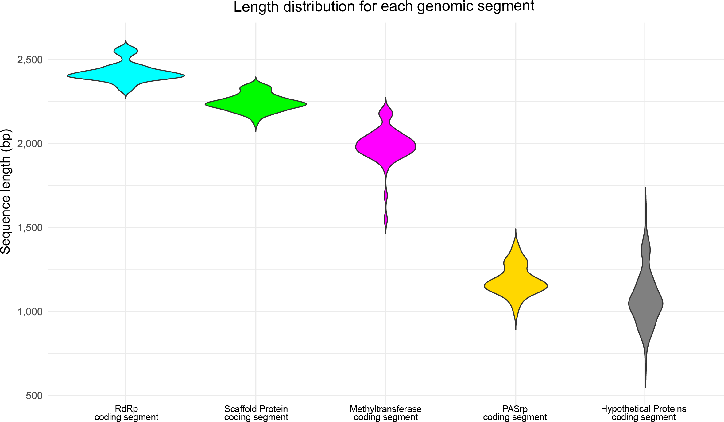 |
| Figure 2 Polymycoviridae. Violin plots showing the length distribution of individual genomic dsRNA segments from viruses classified in the family Polymycoviridae. The core genome segments display relatively conserved size ranges, whereas additional genomic segments (dsRNA extras) exhibit greater length variability among different virus species. |
Proteins
The dsRNAs are associated with a proline-alanine-serine rich protein (PASrp; 18–32 kDa), the principal structural component of polymycovirus ribonucleoprotein complexes (Kanhayuwa et al., 2015, Zhai et al., 2016, Kotta-Loizou and Coutts 2017, Niu et al., 2018). In CcFV1, PASrp is the major structural protein of filamentous capsids (Jia et al., 2017). Whether capsid formation in CcFV1 requires additional viral proteins remains to be determined.
Genome organization and replication
The genomes of polymycoviruses consist of four to eight linear dsRNA segments (Figure 3 Polymycoviridae). Each segment contains a single major open reading frame (ORF), flanked by non-coding regions (NCRs) of variable length. Terminal sequences are conserved between segments of particular viruses but show limited conservation across species. No evidence for extensive sequence complementarity or panhandle structures at the NCRs has been reported.
The conserved core genome of polymycoviruses comprises four dsRNA segments. One of the conserved genomic segments encodes the RNA-directed RNA polymerase (RdRP) responsible for viral genome replication. The RdRP of polymycoviruses belongs to the protein family RdRP_1 (Pfam PF00680) and has three conserved motifs in its catalytic domain, including the conserved GDNQ amino acid motif. Another conserved genomic segment encodes a protein of unknown function, putatively a scaffold protein for replication and virion assembly (Kotta-Loizou and Coutts 2017). This protein is characterized by a conserved N-terminal region, a cysteine-rich, zinc finger-like motif, and is rich in arginine repeats (R-R, R-X-R), which are associated with endoplasmic reticulum (ER) retention signals. The third conserved genomic segment encodes a methyltransferase that is responsible for adding a capping structure at the 5′-termini of the (+) viral RNAs (Kanhayuwa et al., 2015, Kotta-Loizou and Coutts 2017). Polymycovirus methyltransferases display the typical two-domain organization of methyltransferases found across all kingdoms of life, consisting of a methyltransferase catalytic motif and an N-terminal Rossmann-fold domain. The fourth conserved genomic segment encodes the proline–alanine–serine-rich protein (PASrp), which constitutes the principal structural component of polymycovirus ribonucleoprotein complexes and, in CcFV1, the major structural protein of the filamentous capsid. In members of several polymycovirid species, the conserved four-segment core genome is supplemented by additional genomic segments. When present, these extra segments encode proteins with no detectable homology to proteins of known function and are non-homologous among different polymycovirids and do not interfere with taxonomic classification.
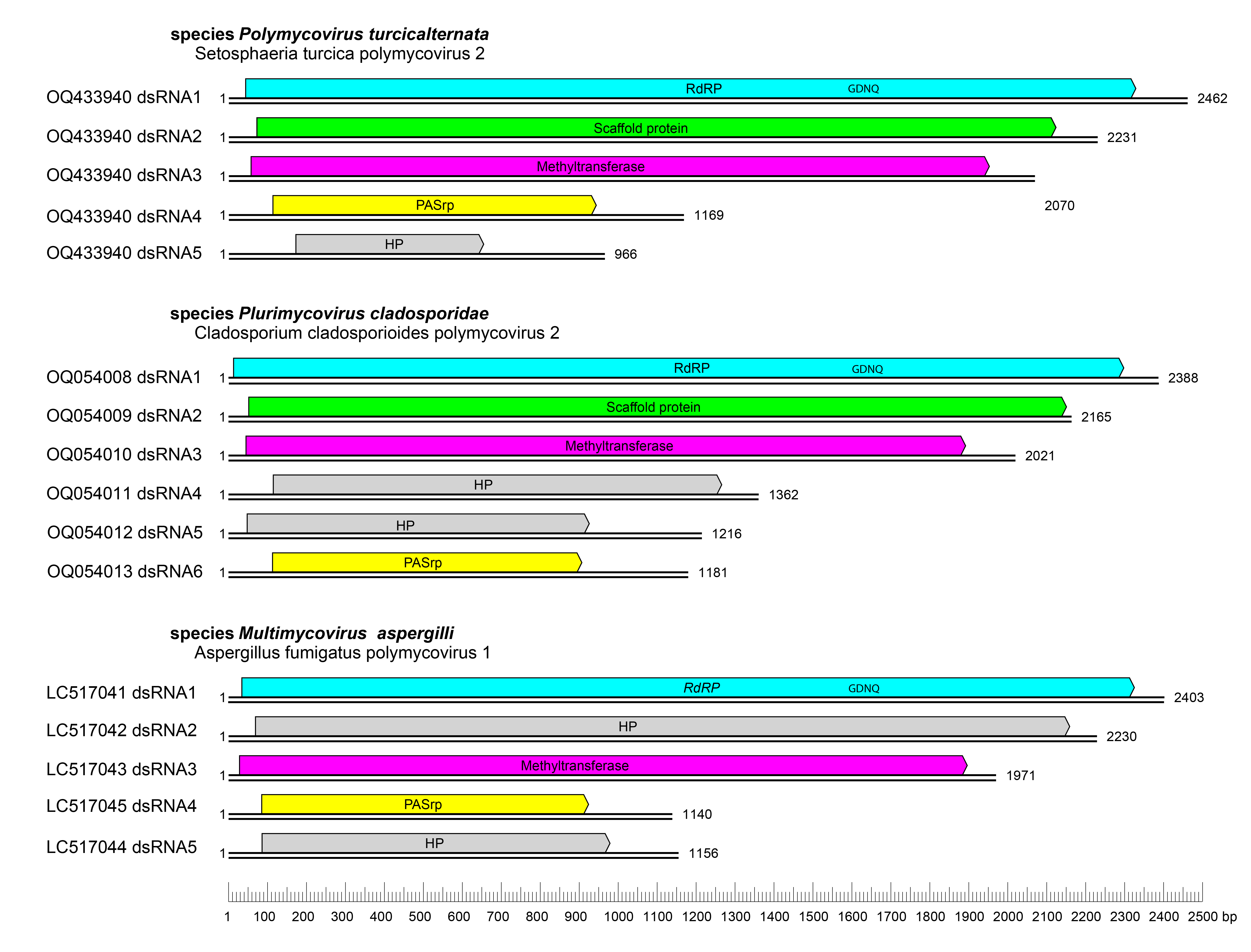 |
| Figure 3 Polymycoviridae. Genome organization of polymycovirids. The genome consists of 1–8 monocistronic dsRNA segments. The ORFs of the core genomic segments are shown in cyan, green, magenta and yellow, whereas ORFs of additional genomic segments (dsRNA extras), which encode hypothetical proteins, are shown in gray. The position of the conserved RdRP catalytic motif (GDNQ) is indicated on dsRNA1. |
Biology
Polymycovirids lack an extracellular phase in their life cycle; they are transmitted intracellularly during cell division and sporogenesis (vertical transmission) and by cell fusion following hyphal anastomosis between compatible fungal strains (horizontal transmission) (Zhai et al., 2016, Filippou et al., 2018). Virions accumulate in the cytoplasm. These viruses were the first dsRNA viruses found to be infectious not only as purified entities but also as naked dsRNA (Kanhayuwa et al., 2015, Jia et al., 2017, Niu et al., 2018). Association with a broad range of host phenotypic alterations has been reported, including changes in pigmentation (Kanhayuwa et al., 2015, Jia et al., 2017, Kotta-Loizou and Coutts 2017, Filippou et al., 2021) and sectoring (Kanhayuwa et al., 2015, Zhai et al., 2016, Kotta-Loizou and Coutts 2017). Polymycovirid infection has also been associated with decreased host growth (Zhai et al., 2016, Jia et al., 2017) and host virulence in plants (Zhai et al., 2016, Jia et al., 2017), insects (Pitaluga et al., 2020) and mammals (Takahashi-Nakaguchi et al., 2020). In contrast, increased sporulation (Filippou et al., 2021), enhanced host growth (Kotta-Loizou and Coutts 2017, Filippou et al., 2021) and elevated host virulence in the greater wax moth (Galleria mellonella) study model (Özkan and Coutts 2015, Kotta-Loizou and Coutts 2017) have also been described. Additional effects include increased host sensitivity to a range of stresses (Sass et al., 2023b, Stevens et al., 2023), to antifungal compounds (Niu et al., 2018, Sass et al., 2023a) and to the bacterium Pseudomonas aeruginosa (Nazik et al., 2021). Although the molecular mechanisms underpinning these phenotype changes remain poorly understood, there is evidence that polymycovirids modulate host carbon, nitrogen, and iron metabolism (Filippou et al., 2021, Patil et al., 2021). Finally, polymycovirids are targeted by the host RNA silencing machinery (Özkan et al., 2017).
Antigenicity
High-affinity, PASrp-specific polyclonal antibodies have been produced in mice and rabbits following immunization with purified virions or recombinant PASrp (Zhai et al., 2016, Jia et al., 2017).
Derivation of names
Multimycovirus: variant of the original genus name Polymycovirus, in which the prefix poly is replaced by multi, denoting the presence of multiple genomic segments
Polymycoviridae and Polymycovirus: from the Greek poly meaning “many”, referring to the multi-segmented dsRNA genome, and myco, meaning “fungus”, indicating the fungal hosts
Plurimycovirus: variant of the original genus name Polymycovirus, in which the prefix poly is replaced by pluri, denoting the presence of multiple genomic segments.
Genus demarcation criteria
Genus assignment is primarily based on phylogenetic analysis of RdRP amino acid sequences. Viruses are classified into a given genus when they cluster within a well-supported monophyletic clade corresponding to that genus in RdRP-based phylogenetic trees.
Relationships within the family
The family Polymycoviridae includes three genera, Polymycovirus, Multimycovirus and Plurimycovirus, each corresponding to a well-supported monophyletic clade based on RdRP amino acid sequence phylogeny (Figure 4 Polymycoviridae).
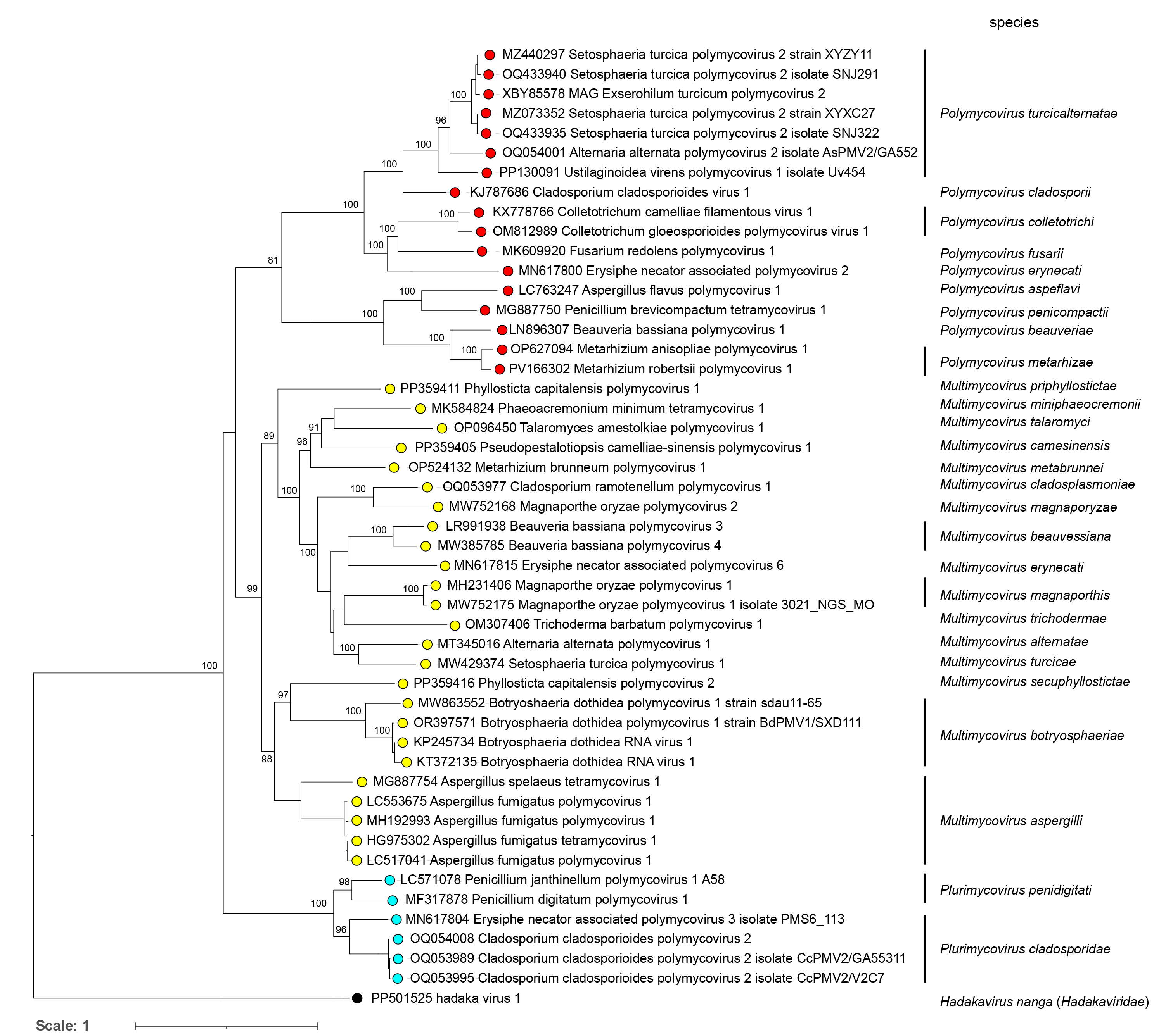 |
| Figure 4 Polymycoviridae. Phylogenetic analyses of the RdRP amino acid sequences of members of the family Polymycoviridae. The sequences were downloaded from NCBI on April 10th, 2025. MAFFT v7.471 was used for multiple sequence alignment. The ML phylogenetic tree was constructed using IQ-TREE v1.6.11, which calculates the best evolutionary model (LG+F+I+G4). The three genera are indicated. |
Relationships with other taxa
The family Polymycoviridae is assigned to the phylum Pisuviricota, kingdom Orthornavirae, within the realm Riboviria, and is classified in the class Mycopleornaviricetes and the order Xenadelphovirales, together with the phylogenetically related family Hadakaviridae. Phylogenetic analyses based on the amino acid sequences of the RdRP, the hallmark protein of non-reverse-transcribing viruses across Riboviria, support the close evolutionary relationship between Polymycoviridae and Hadakaviridae. Members of both families cluster within a well-supported monophyletic clade that is clearly separated from other families in the phylum Pisuviricota, including Picornaviridae, Astroviridae, and Partitiviridae (Figure 5 Polymycoviridae). Hadakaviridae comprises +ssRNA viruses, whereas Polymycoviridae members possess dsRNA genomes. The RdRPs of polymycoviruses are related to those of several +ssRNA virus families, including Astroviridae, Caliciviridae and Picornaviridae, as well as to those of the dsRNA family Partitiviridae. Notably, polymycovirus RdRPs conserve a GDNQ motif in the palm domain, a feature more commonly associated with −ssRNA viruses, rather than the canonical GDD motif typical of most dsRNA and +ssRNA viruses. Together with the variable presence of capsid-like structures, these features position polymycoviruses as evolutionary intermediates between dsRNA and +ssRNA viruses, as well as between encapsidated and capsidless RNA viruses (Kanhayuwa et al., 2015, Oliveira et al., 2025).
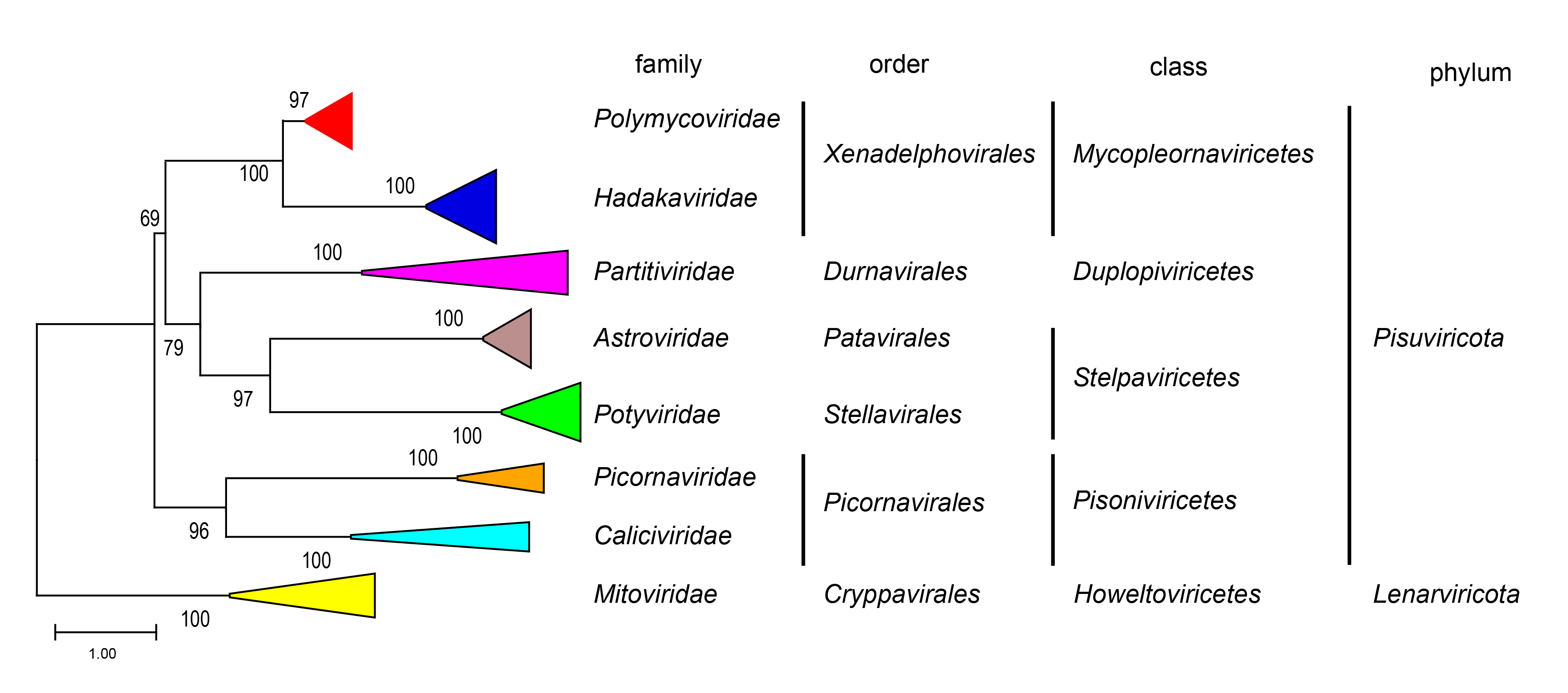 |
| Figure 5 Polymycoviridae. Phylogenetic analyses of the RdRP amino acid sequences of viruses in the families Polymycoviridae, Hadakaviridae, and other viral families within the phylum Pisuviricota. The hadakaviruses and polymycoviruses sequences were downloaded from NCBI on April 10th, 2025, and representative sequences of the viral families within the phylum Pisuviricota were selected from the VMR_MSL40.v2 list. MAFFT v7.471 was used for multiple sequence alignment. The ML phylogenetic tree was constructed using IQ-TREE v1.6.11, which calculates the best evolutionary model (LG+F+R5). |
Related, unclassified viruses
These viruses cannot yet be formally classified because their genomes are not fully sequenced and some segments of the core genome are missing, preventing reliable taxonomic assignment.
| Virus* | Accession number |
| Sclerotinia sclerotiorum tetramycovirus 1 | dsRNA1: MF444217 dsRNA2: MF444218 dsRNA3: MF444219 |
| Erysiphe necator associated polymycovirus 5 | dsRNA1: MN617811 |
| Erysiphe necator associated polymycovirus 8 | dsRNA1: MN617819.1 dsRNA2: MN617820 |
| MAG Exserohilum turcicum polymycovirus 2 | dsRNA1: PP926250 |
| Plasmopara viticola lesion associated polymycovirus 3 | dsRNA1: MN557034 |
| Erysiphe necator associated polymycovirus 6 | dsRNA1: MN617815 dsRNA2: MN617816 dsRNA3: MN617817 |
| Beauveria bassiana polymycovirus 2 | dsRNA1: LN896311 dsRNA2: LN896312 dsRNA3: LN896313 |
| MAG Exserohilum turcicum polymycovirus 1 | dsRNA1: PP926255.1 |
| Plasmopara viticola lesion associated polymycovirus 1 | dsRNA1: MN557029 dsRNA2: MN557030 |
| Erysiphe necator associated polymycovirus 4 | dsRNA1: MN617808 |
| Plasmopara viticola lesion associated polymycovirus 5 | dsRNA1: MN557036 dsRNA2: MN557037 dsRNA3: MN557038 |
| Alternaria tenuissima virus | dsRNA1: KP067914 |
| Alternaria sp. FA0703 | dsRNA2: FJ595830 dsRNA5: FJ595831 dsRNA6: FJ595832 |
| Melampsora lini | dsRNA3: X64371 |
| mycovirus M7 | dsRNA1: MN418003 |
| Plasmopara viticola lesion associated polymycovirus 2 | dsRNA1: MN557032 dsRNA2: MN557031 dsRNA4: MN557033 |
| Plasmopara viticola lesion associated polymycovirus 4 | dsRNA1: MN557035 |
* Virus names are not official ICTV designations.